Product info
- Product name:Dry Eye Detector
- Product code:C260225003
Packaging Specifications
- Outer Packaging Size:0.56 × 0.39 × 0.33 m
- Gross Weight:15 kg
- Net Weight:13 kg
- Volume:0.07
Description
OCULAR SURFACE ANALYZER SERIES
PROFESSIONAL DRY EYE + DIGITAL SLIT LAMP LIGHT SOURCE
AUTO QUANTITATIVE PARAMETER
model
Optional slit lamp
Digital collector
Uniform halo
TMH
NIBUT
Lipid Layer Static
Lipid Layer Dynamic
Incomplete blinking
Red Eyes
Gland opening
Meibo
Corneal Staining
Mite function
Manual
Automatic
CCD
Yes
NIBUT
Infrared photography:
Using infrared as the light source, it does not stimulate tear secretion, making it more comfortable and the results more realistic.
Visible light shooting:
Penetrating white light technology allows for clearer viewing of the tear stream, making it easier to observe its continuity.
TMH
Infrared photography:
Using infrared as the light source, it does not stimulate tear secretion, making it more comfortable and the results more realistic.
Visible light shooting:
Penetrating white light technology allows for clearer viewing of the tear stream, making it easier to observe its continuity.
LIPID LAYER DYNAMIC
By using a uniform mask projection and recording automatic progress analysis, the entire range of the lipid layer can be clearly observed, and real-time changes in lipid layer thickness can be observed during each eye opening period.
Using professional AI algorithms, quantitative analysis of lipid layer thickness measurement can be accurate to 1nm.
BLINKING
Automatic recognition, statistics, and playback
In the lipid layer dynamic analysis project, simultaneously automatically identify and record incomplete blink data.
GLAND OPENING
High definition photography
Can clearly obtain the overall shape of the eyelid margin images of subtle changes in glandular opening.
MEIBO
Professional infrared imaging can clearly observe the morphology of meibomian gland
AI image enhancement:
Supports automatic recognition, enhances gland contrast, and automatically calculates the proportion of meibomian gland loss.
LIPID LAYER STATIC
High precision
Adopting uniform mask projection, it can fully present the true color and shape of the lipid layer
Using automatic AI algorithms, the measurement of lipid layer thickness can be accurate to 10nm.
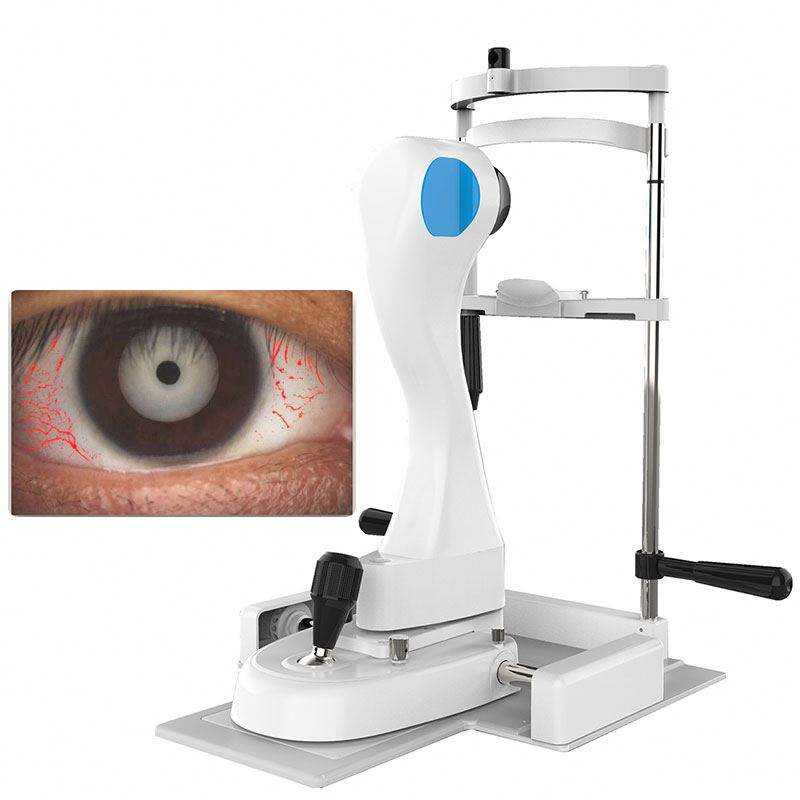
RED EYES
Conjunctiva, ciliary, two modes
Multidimensional analysis of ocular surface congestion in subglands
Provide more basis for diagnosis.
STAINING
Professional yellow filter
Make corneal fluorescein sodium staining images clearer
no comments