Product info
- Product name:50KW 630mA Dual-column 4-dimensional flatbed DR (Digital Radiography) Xray Machine
- Product code:CCX56A
Packaging Specifications
- Outer Packaging Size:2.35 × 1.45 × 0.92 m
- Gross Weight:465 kg
- Net Weight:314 kg
- Volume:3.13
- Outer Packaging Size:1.9 × 1.06 × 1.13 m
- Gross Weight:210 kg
- Net Weight:144 kg
- Volume:2.28
- Outer Packaging Size:2.33 × 0.81 × 0.62 m
- Gross Weight:168 kg
- Net Weight:121 kg
- Volume:1.17
- Outer Packaging Size:0.84 × 0.58 × 1.03 m
- Gross Weight:90 kg
- Net Weight:80 kg
- Volume:0.5
Description
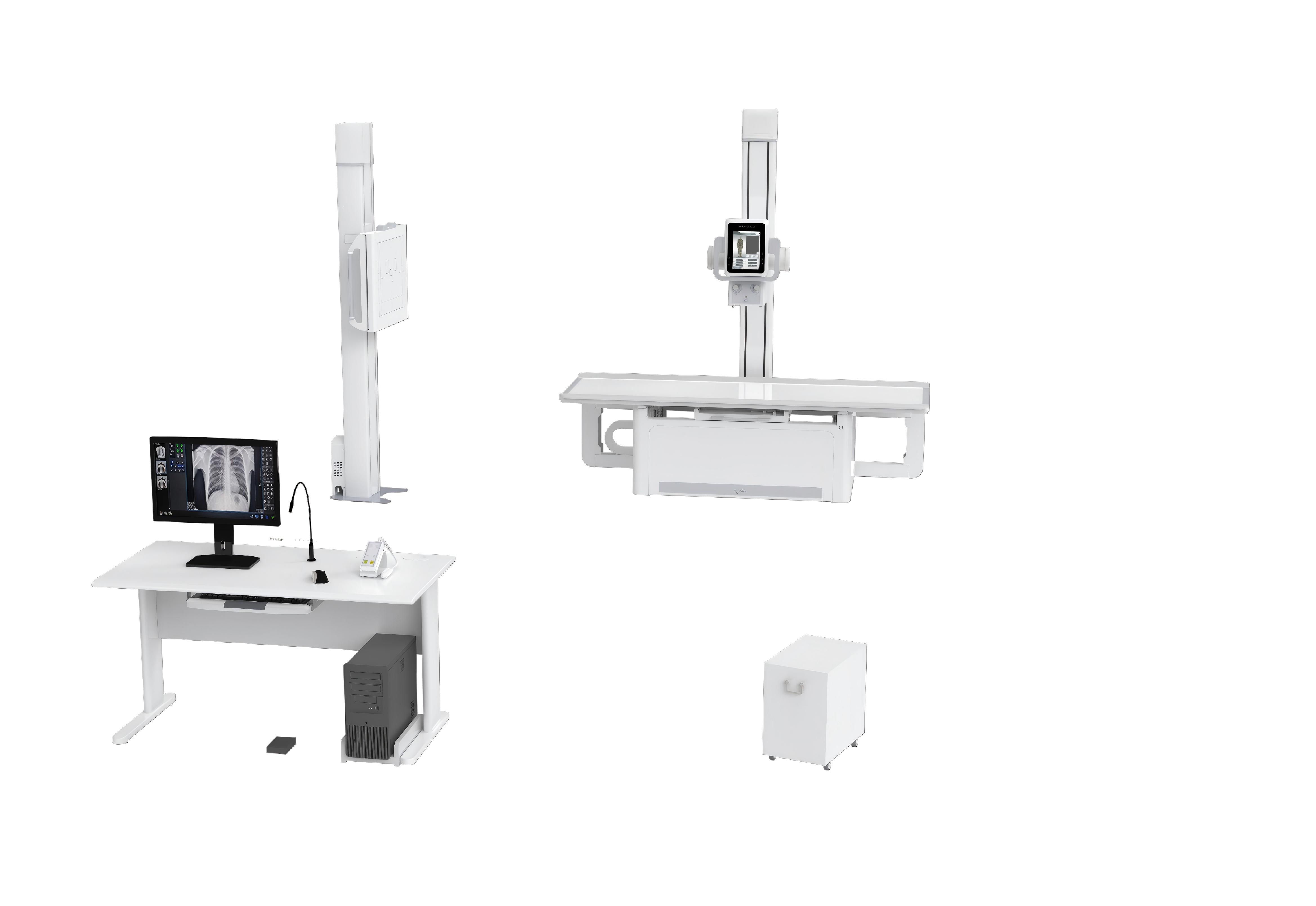
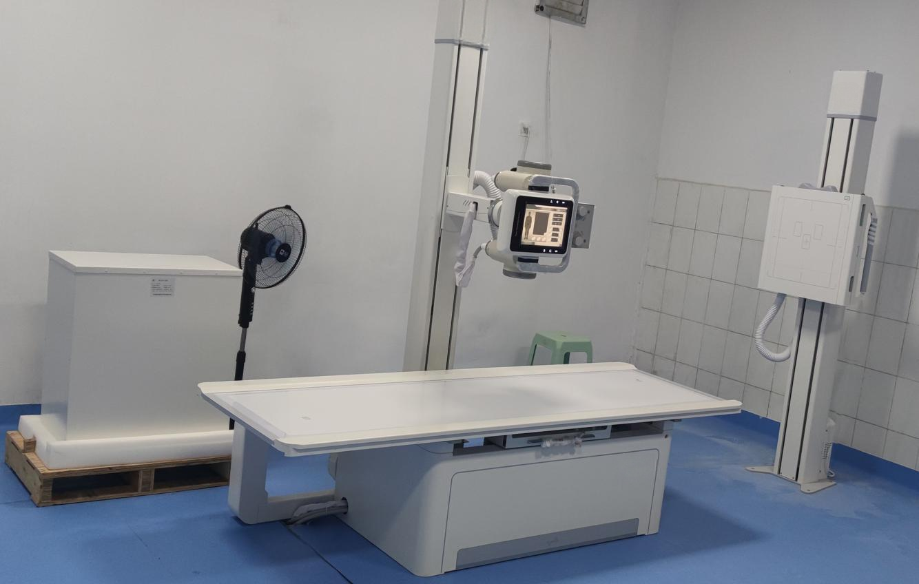
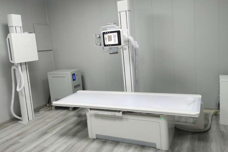
CCX56A 50KW 630mA Dual-column 4-dimensional Flatbed DR X-ray Machine
Core Overview
CCX56A is a floor-mounted dual-column 4-dimensional flatbed digital radiography (DR) X-ray machine with core configurations of 50KW high-frequency generator, 630mA tube current and 17*17inch FPD, matching a 4-way floating table. It features flexible mechanical movement, humanized table-side touch control, high-definition imaging and a powerful digital workstation, supporting full-body radiographic examinations for all human parts. Suitable for radiology departments, orthopedics, wards, emergency rooms, operating rooms, ICUs, fever clinics and physical examination centers, it realizes efficient, easy and low-radiation digital radiography, with complete image acquisition, processing and output capabilities, meeting the diverse clinical imaging needs of medical institutions.
Core Clinical Application
Applicable to radiology department, orthopedics, ward, emergency room, operating room, ICU, fever clinic, children's department and physical examination/ diagnostic centers; supports radiographic examinations of all human body parts including head, limbs, chest, spine, lumbar vertebrae and abdomen, adapting to routine diagnosis, emergency treatment, intraoperative testing and large-scale physical examination scenarios.
Key Mechanical Movement & Structural Parameters
1. Flexible Multi-Dimensional Movement
- Table: 4-way floating table, longitudinal travel 1000mm, transverse travel 260mm, size 2250×810mm, maximum load capacity 200kg
- Tube column/head: Tube column longitudinal travel 1300mm, rotation -180°~+180°; tube head rotation -90°~+90°
- Detector mechanism: Bucky stand equipped, detector mechanism center travel range 1500mm
- Other movements: Support tube up/down, Bucky stand detector up/down, spherical column longitudinal moving, tube rotation/swing around axis
2. Grid Configuration
- Standard 2 sets of removable high-density wire grids (1m/1.8m focal length), specification 18inch*18inch, 103LP, 10:1; removable for children/low-dose part shooting to reduce radiation.
Core Technical Specifications
1. Power & High-Frequency HV Generator
- Power supply: 380V±38V, 50Hz±10Hz, three phase
- Generator: 50KW output power, 500KHz inverter frequency; tube voltage 40kV-150kV (1kV step), tube current 10mA-630mA, mAs 0.1-630mAs
- Collimator/Filter: Illumination brightness ≥200Lux; filter ≥1mmAL
2. X-ray Tube (Model XQG50)
- Dual focus: 1.2mm (big)/0.6mm (small), input power 74kW/30kW
- Target surface 12°, rotating anode speed 8400rpm; anode thermal capacity 300KHU, heat dissipation 750W, total tube capacity 1300KHU
- Special filter: 0.9mm AL/75KV, work voltage 40-150KV
3. Flat Panel Detector (FPD)
- Material: A-Si(Amorphous Silicon)-CSI
- Active area: 430(H)×430(V)mm (17*17inch), pixel matrix 3072×3072, pixel pitch 139μm
- Performance: Spatial resolution 3.7LP/mm, 16bit A/D conversion, ensuring high-definition imaging and subtle tissue detail capture
Professional Digital Workstation & Software Functions
Equipped with a 19〞imported high-resolution medical LCD monitor (brightness/contrast up to 1000NIT, far higher than ordinary 400NIT LCD screens), the workstation integrates registration, image acquisition, processing, output and system protection with rich functions, and supports DICOM3.0 standard:
- Registration: Auto registration + DICOM Worklist SCU, simplifying input and improving work efficiency
- Image Acquisition: Automatic window adjustment, cropping and transmit, realizing one-click efficient acquisition
- Image Processing: Rich tools including tissue equilibrium, W/L adjustment, Gamma correction, noise reduction, sharpening, pseudo color, multi-angle rotation/mirror, and length/angle/area measurement; built-in optimization algorithms for different human physiological structures/parts
- Image Output: DICOM3.0 format support, compatible with PACS/HIS/RIS system connection; support film printer output, paper report (up to 3 reference images), and USB/DVD image copy
- System Support & Protection: Built-in operation manual, microphone + remote exposure control (outside X-ray room), infrared anti-misoperation facilities, overloading system with error code alarm and self-protection design
- Predefined Programs: More than 200,000 anatomical programs for standardized and quick exposure parameter selection
Core Product Advantages
1. Humanized Table-side Touchable Control System (Synchronized with Workstation)
- Realize cross-room exam creation (X-ray/control room), no repeated settings, convenient for routine and emergency diagnosis
- Support post-exposure image preview, real-time SID stretching display and exposure parameter setting
- Human graphic body part selection, collimator effective area adjustment and digital display of tube head rotation angle
- 3D model positioning demonstration, gravity sensor automatic rotation for precise positioning
- Emergency exam access: Shoot first then register with automatic record, saving precious emergency time
2. Flexible 4-dimensional Mechanical Movement & High Adaptability
- 4-way floating table + multi-directional movement of tube/detector, meeting various clinical positioning needs for different parts and postures
- Tube column/head wide-angle rotation, adapting to special position radiography; 200kg large table load capacity, suitable for all types of patients
- No need to modify on-site facilities, easy to install and use in various clinical departments
3. Double Radiation Protection & High Imaging Quality
- 2 sets of standard removable high-density wire grids, effectively filtering scattered rays to improve image contrast; removable for children/low-dose parts to reduce radiation exposure for doctors and patients
- 50KW high-frequency generator + 16bit high-resolution FPD, ensuring stable radiation output and high-definition, detail-rich imaging; 1kV step tube voltage adjustment for precise dose control
4. Efficient & Integrated Workflow
- Automatic acquisition/processing/transmit functions, reducing manual operation and improving examination efficiency
- DICOM3.0 full compatibility, realizing seamless connection with hospital information systems, facilitating image sharing, archiving and remote diagnosis
- Over 200,000 predefined anatomical programs, realizing one-click parameter selection and standardized radiography, suitable for large-scale physical examinations and high-flow outpatient scenarios
5. Reliable System Protection & Easy Operation
- Infrared anti-misoperation facilities, overloading alarm and self-protection design, ensuring safe equipment operation
- Intuitive graphical operation interface, built-in operation manual, and table-side/remote dual control mode, reducing training costs and lowering operation threshold for medical staff
no comments