
Product info
- Product name:Portable Full Digital Ultrasound Diagnostic Scanner with Multiple Probes
- Product code:CCBK7
Packaging Specifications
- Outer Packaging Size:0.42 × 0.4 × 0.21 m
- Gross Weight:8.5 kg
- Net Weight:8 kg
- Volume:0.04
Description
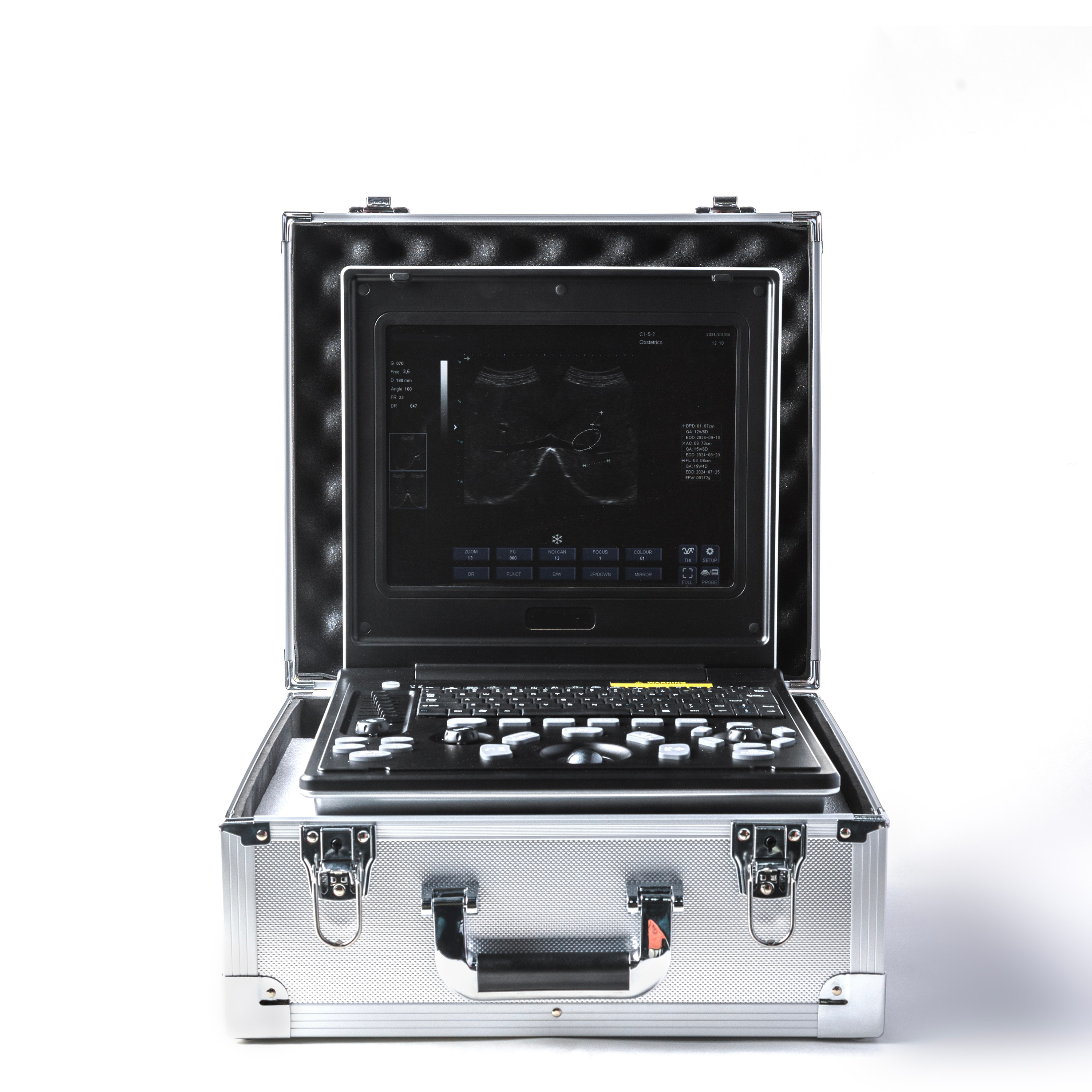
Portable Full Digital Ultrasound Diagnostic Scanner
The CCBK7 portable full digital ultrasound diagnostic scanner is a lightweight notebook-type ultrasonic imaging device for general clinical examinations. Adopting advanced full digital imaging technology and multi-mode scanning design, it supports multi-department routine ultrasound diagnosis. With built-in battery and dual-probe compatibility, it meets stationary clinical diagnosis and emergency mobile scanning needs for hospitals, clinics and field medical services.
Features
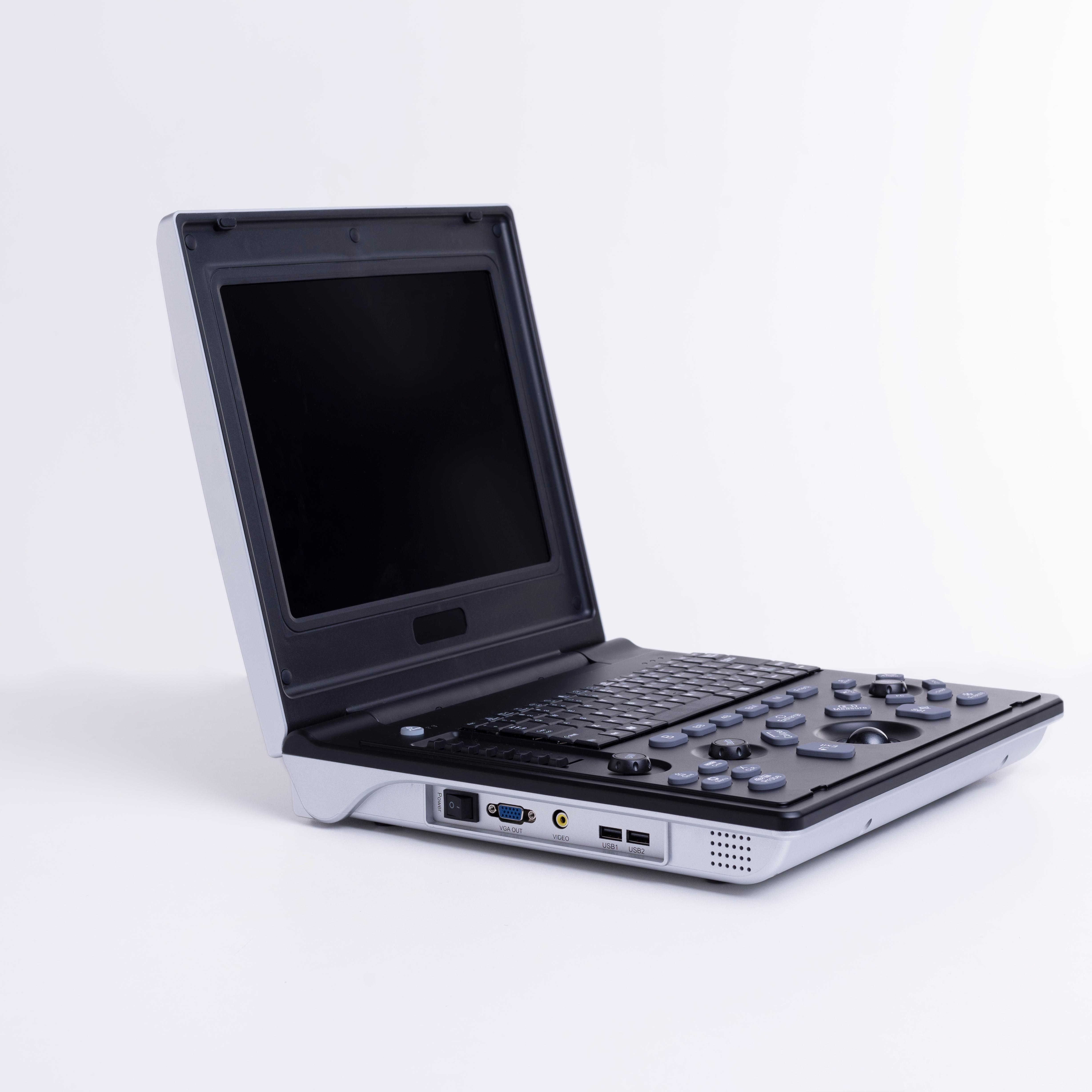
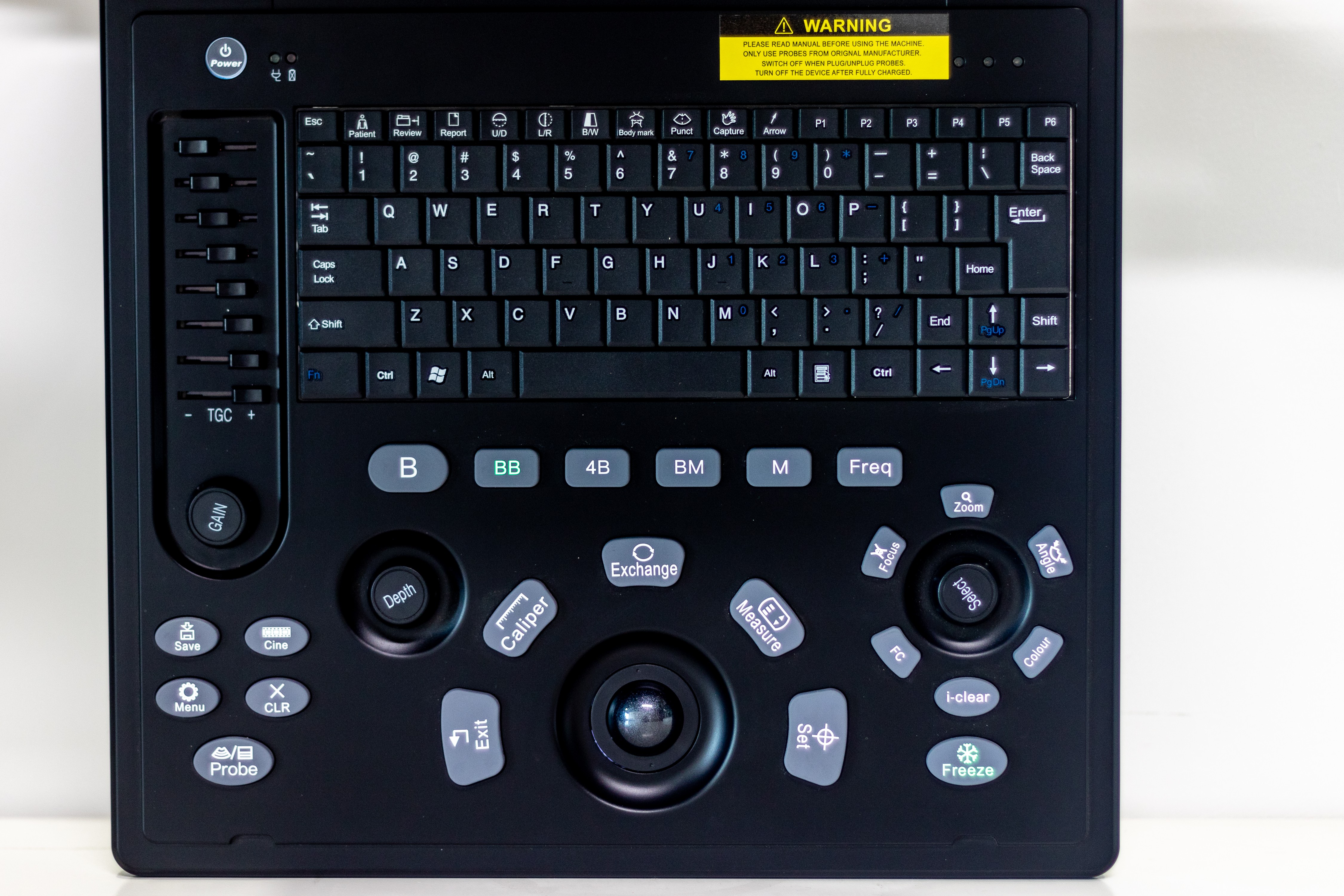
● Ultra-portable notebook design with rechargeable battery for mobile use
● 12-inch high-definition LED LCD display for clear real-time imaging
● Professional full digital imaging processing system for stable image output
● Multiple clinical imaging modes: B, B+B, B+M, M, 4B mode
● Automatic probe identification for fast mode switching
● One-key intelligent image optimization for efficient operation
● Built-in cine playback function for dynamic image review
● Local image storage and USB external expansion storage support
● Adjustable scanning angle and multi-level image magnification
● Customizable puncture guide line (adjustable angle and position)
● Comprehensive measurement tools and standardized report generation functions
Clinical Applications
Suitable for:
● Routine abdominal organ examination
● Obstetric and gynecological scanning
● Cardiac structure and function examination
● Urological system diagnosis
● Pediatric ultrasound examination
● Superficial small organ imaging
● Musculoskeletal tissue detection
● Emergency bedside and mobile ultrasound diagnosis
Technical Specifications
Display
● 12-inch LED LCD screen
● Resolution: 1024 × 768
Imaging Modes
● B / B+B / B+M / M / 4B
Image Functions
● Image smoothing and sharpening adjustment
● Tissue harmonic imaging processing
● Gamma correction optimization
● False color imaging processing
● Adjustable dynamic range
● Multi-point focus adjustment
Storage Performance
● Cine playback capacity: ≥256 frames
● Built-in image storage: ≥300 frames
● USB external storage expansion
● Fast image saving (≤9 seconds)
Measurement Functions
General Measurement
● Distance / Circumference / Area / Volume
Cardiac Measurement
● Left atrium / Right atrium / Left ventricle / Right ventricle / Aorta
Obstetric Measurement
● Gestational sac / BPD / Head circumference / Abdominal circumference / Femur length / Amniotic fluid volume
● Automatic gestational age calculation and fetal weight estimation
Gynecological Measurement
● Uterus / Cervix / Endometrium / Ovary / Follicle
Urology Measurement
● Kidney / Prostate / Testis / Epididymis
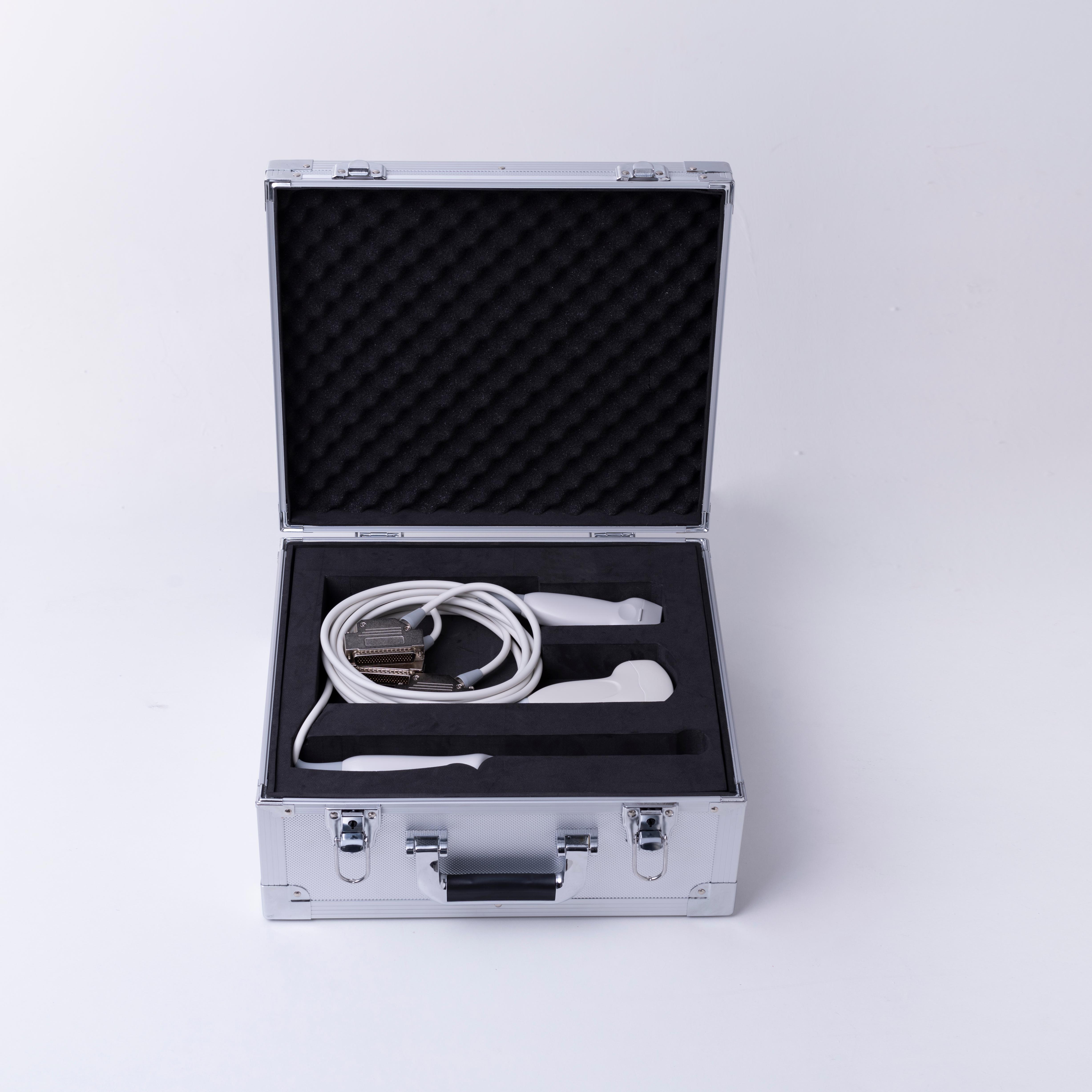
Probe Options
● 3.5MHz Convex probe
● 6.5MHz Intracavity probe
● 7.5MHz Linear probe
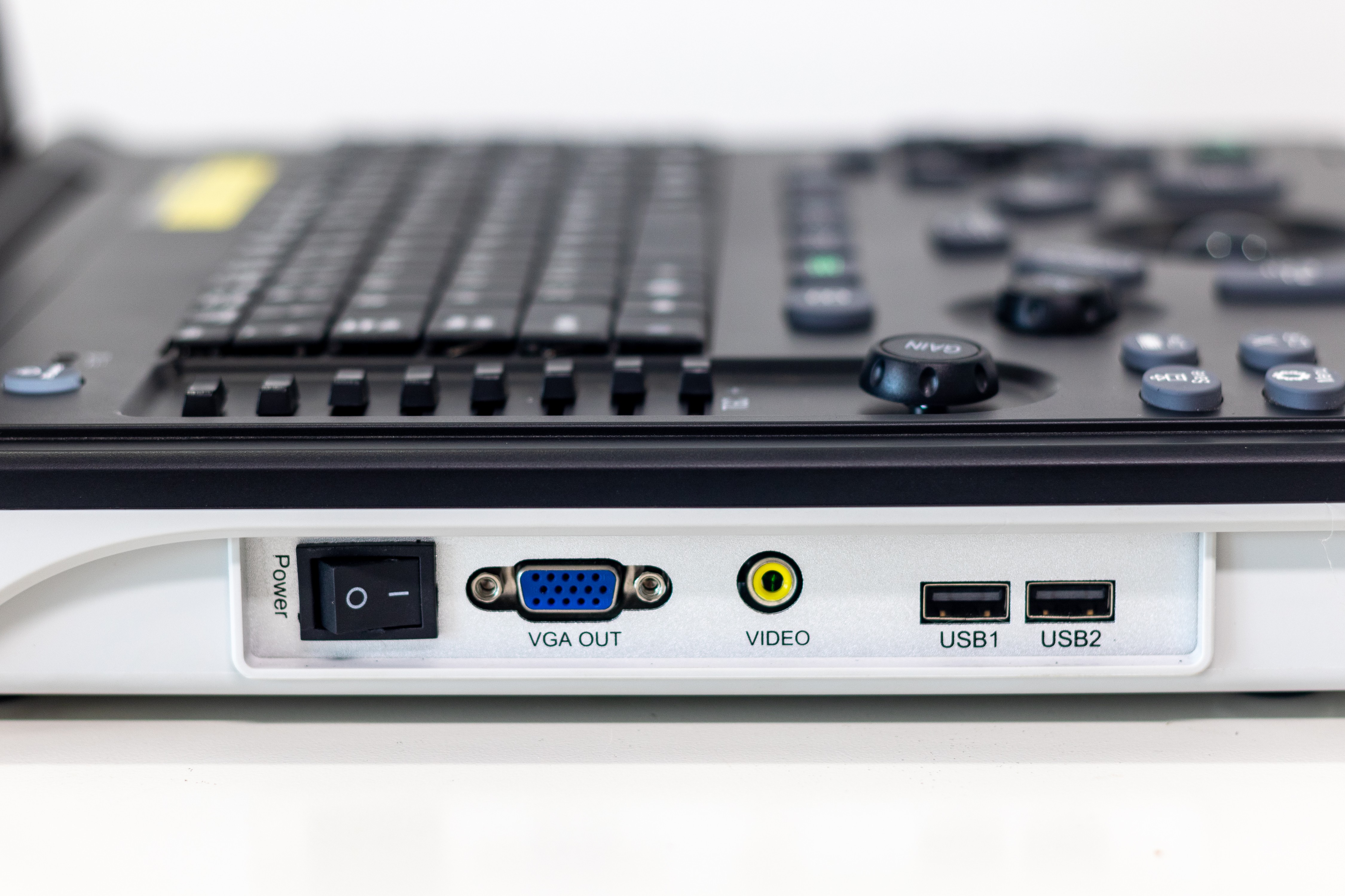
Interfaces
● VGA output interface
● USB 2.0 port
● A/V interface
● Dual probe simultaneous connection
Product Advantages
● Lightweight portable design adaptable to ward, bedside and mobile scenarios
● Full-featured routine ultrasound solution for multi-department diagnosis
● Replaceable probes to meet diverse clinical scanning requirements
● Preset examination programs for simple and efficient operation
● Cost-effective and reliable for clinics, hospitals and mobile medical services
no comments