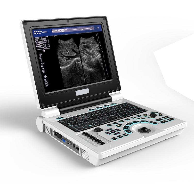
Product info
- Product name:B&W Portable Ultrasound Scanner
- Product code:CCBF30B
Out of stock.
Request a Quote
Packaging Specifications
Core Parameters
Description
Ultrasound Scanner Specifications
Main Specifications
Scanning Mode
- Electronic scanning
Display Modes by Probe Type
- T35R60GB Convex Array Probe: B, B/B, 4B, B+M, M
- T50R20GB Micro Convex Probe: B, B/B, B+M, M
- T65R13GB Cavity Probe: Only B
- T75L40GB Linear Array Probe: Only B
Image Magnification
- T35R60GB Convex Array Probe (B Mode, Real-Time): ×0.8, ×1.0, ×1.2, ×1.5, ×1.8, ×2.0
- Display Penetration Depth: ×0.8, ×2.0
- T65R13GB Cavity Probe: ×0.8, ×1.0, ×1.2, ×1.5
M Mode
- 8 selectable scanning speeds
- Note: Measurement error increases at heart rates over 100 bpm
Zoom
- Local ×2.0 magnification
Dynamic Range
- 0–120dB adjustable
Frequency
- T35R60GB Convex Array Probe: 2.5MHz / 3.0MHz / 3.5MHz / 4.0MHz / 5.0MHz (five-period conversion)
- T65R13GB Cavity Probe / T75L40GB Linear Array Probe: 5.5MHz / 6.0MHz / 6.5MHz / 7.0MHz / 7.5MHz (five-period conversion)
Focus Position
- Sending: 1, 2, 3, and 4-segment dynamic electronic focusing (focus movable for 1- and 2-segment focus)
- Receiving: Real-time dynamic focus
Measuring Function
- General: Distance, Circumference/Area (ellipse method, loci method), Volume, Heart Rate
- Obstetrics: Gestational Weeks (BPD, GS, CRL, FL, HC, AC), Expected Date of Confinement, Fetal Weight, etc.
Annotation Function
- Hospital name, Patient’s name, Age, Gender
- 64 body marks (with probe position)
- Full-screen character annotation
- Real-time clock display
Puncture Guide
- The 3.5MHz convex array probe can display a puncture guide line in B mode.
Gain Control
- 8-segment TGC and overall gain adjustable separately.
Image Polarity
- Left/right flip, black/white flip, up/down flip.
Permanent Storage
- ≥128 images.
Image Playback
- Series playback or check one by one.
Image Processing
- γ correction, Frame correlation, Point correlation, Line correlation, Digital filtering, Digital Edge Enhancement, Pseudo Color Processing, etc.
USB Interface
- Only for U-disk image storage; format: BMP.
Output Interface
- SVGA video output (connect to color display, workstation, etc.)
- PAL video output (connect to display, video image recorder, etc.)
Probe Functions
Convex Probe
- Frequency: 2.0MHz, 2.5MHz, 3.5MHz, 4.0MHz, 5MHz
- Depth: 16 levels (126, 142, 158, 173, 189, 197, 205, 213, 221, 229, 237, 245, 253, 261, 269, 307mm)
- Applications: Abdominal, Obstetrics, Gynecology, Regional nerve block, Procedure and Biopsy guidance
Linear Probe
- Frequency: 5.5MHz, 6.5MHz, 7.0MHz, 7.5MHz, 9.0MHz
- Depth: 16 levels (35, 42, 49, 62, 69, 76, 83, 90, 97, 104mm)
- Applications: Breast, Small parts, Nerve, Vascular, Venous access, Musculoskeletal, Biopsy guidance
Trans-vaginal Probe
- Frequency: 5.0MHz, 5.5MHz, 6.5MHz, 7.0MHz, 7.5MHz
- Depth: 16 levels (62, 69, 76, 83, 90, 97, 104, 111, 125, 139mm)
- Applications: Obstetrics, Gynecology
Micro-convex Probe
- Frequency: 4.0MHz, 4.5MHz, 5.0MHz, 6.5MHz, 7.0MHz
- Depth: 16 levels (62, 69, 83, 97, 111, 125, 139, 152, 166, 180mm)
- Applications: Pediatric heart, basic function
no comments