Product info
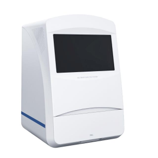
- Product name:Chemiluminescence Imager
- Product code:C26102206
Out of stock.
Request a Quote
Packaging Specifications
Core Parameters
Description
All-in-One Chemiluminescence Gel Imaging System
Instrument Introduction
This all-in-one chemiluminescence gel imaging system adopts computer-free integrated design and is equipped with a 15.6-inch high-resolution intelligent touch screen system. The whole experimental process can be completed directly through multi-directional touch operation. The system integrates four core functional modules: chemiluminescence imaging, protein gel imaging, nucleic acid gel imaging and gel cutting system, which covers the imaging requirements of various experimental scenarios in one stop. The instrument is suitable for routine detection in molecular biology, cell biology, proteomics and other fields.
Detection Principle
Protein Gel Imaging: For protein samples stained with Coomassie brilliant blue, silver stain or fluorescent protein dyes (such as SYPRO Ruby), the transmission or reflection light signals are collected under the irradiation of high-brightness LED white light to realize high-resolution imaging of protein gels.
Nucleic Acid Gel Imaging: Nucleic acid dyes such as ethidium bromide (EB) bind to DNA/RNA and emit fluorescence under ultraviolet excitation. High-sensitivity sensors capture fluorescence signals to obtain images of nucleic acid bands in gels.
Chemiluminescence Imaging: Horseradish peroxidase (HRP) catalyzes the substrate to generate chemiluminescence signals. The luminescence signals are directly captured without excitation light, so as to realize high-sensitivity and low-background detection such as Western Blot.
Application Scope
Nucleic Acid Gel Imaging: Analysis of DNA/RNA agarose gel electrophoresis bands, PCR product detection, detection of nucleic acid probe hybridization signals, etc.
Protein Gel Imaging: Protein detection and molecular weight identification after Coomassie brilliant blue staining, silver staining, etc.
Chemiluminescence Detection: Western Blot, Southern/Northern Blot, dot blot, enzyme activity detection, etc.
Instrument Features
- Multi-function integration: Integrate three shooting modes of ultraviolet, white light and chemiluminescence, covering the full-scenario imaging requirements of nucleic acid, protein and chemiluminescence. No equipment replacement is required, which greatly improves the experimental efficiency.
- Ultra-high resolution and sensitivity: Equipped with a CMOS camera with ultra-high resolution and sensitivity and an original lens with high transmittance to ensure high-quality imaging results. The dynamic range of the camera is 16000:1 with an extremely wide signal measurement range. The transmittance of the optical lens to light in the wavelength range of 400~900nm is ≥85% to avoid attenuation of weak signals. The two core indicators guarantee the ultra-wide dynamic measurement range and ultra-high sensitivity of the system.
- Intelligent exposure mode: Built-in intelligent exposure algorithm, which automatically judges the signal intensity and recommends the optimal exposure time to avoid overexposure or underexposure. It is suitable for detection with wide dynamic range such as chemiluminescence, and can intelligently mark overexposed positions.
- Convenient and efficient operation: Adopt computer-free all-in-one design, equipped with 15.6-inch high-definition touch screen with sensitive touch and support for multi-directional operation. Built-in intelligent operation software with pre-set templates for various experimental scenarios, enabling beginners to get started quickly.
- Image processing function: Built-in professional image processing tools. It supports image rotation, cropping, splicing, brightness and contrast adjustment to ensure the original image reaches the best analysis state.
- Multiple protection safety design: Ultraviolet interlock protection device, anti-corrosion and high temperature resistant instrument shell.
Technical Parameters
- Display: 15.6-inch capacitive touch screen
- Shooting Field of View: Effective field of view 150mm×150mm for blot membrane / protein gel / nucleic acid gel
- Camera: 9 million pixels; effective area 11.28mm×11.28mm; dark current <0.0005e-/p/s; camera bit depth 16bit (65536 gray values); camera cooling temperature -25℃@room temperature (22℃)
- Pixel Size: 3.76μm×3.76μm
- Image Resolution: 3000×3000 (9 million pixels)
- Pixel Binning: 1×1Bin, 2×2Bin, 3×3Bin, 4×4Bin, 6×6Bin, 8×8Bin
- Lens: Aperture F0.95, lens transmittance ≥85%
- Filter: Narrowband filter 590nm, bandwidth 10nm±2nm
- White Light Source: LED uniform light strip, symmetrically distributed on both sides
- UV Transilluminator: 6~10W (single lamp tube), service life 3000h, wavelength 302nm, ballast efficiency >80%
- External Computer: Windows10 operating system, 6th generation i5, 16G RAM, 256G SSD
- External Interfaces: USB2.0×2, USB3.0×2, Ethernet port×1
- Working Voltage: AC220V 50Hz
- Dimension: 460mm×470mm×715mm
- Weight: 36.1kg
- Product Power: ≤90W
no comments