Packaging Specifications
Core Parameters
Description
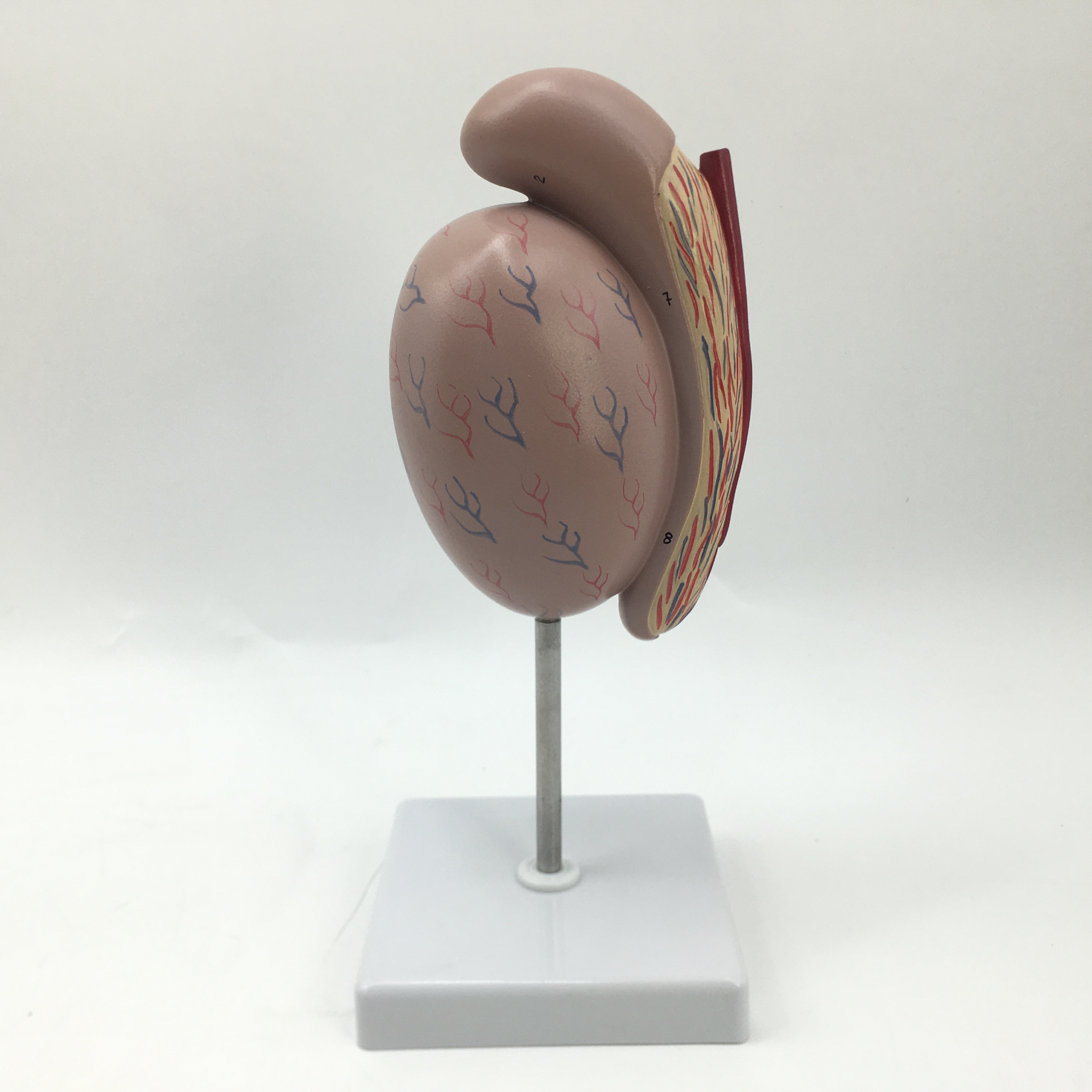
3.5x Magnified Testis Model
Product Description
This model is magnified 3.5 times the size of a normal male testis. It displays the medial surface and sagittal section of the testis, and fully illustrates internal anatomical structures including efferent ductules, tunica albuginea, vaginal cavity, visceral layer of tunica vaginalis, seminiferous tubules, vas deferens and rete testis.
no comments