Product info
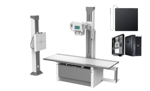
- Product name:65KW 800mA Floor-mountedDigital Radiography System
- Product code:CCX65DR-P
Out of stock.
Request a Quote
Packaging Specifications
Core Parameters
Description
Introduction
Floor-mounted Digital X-ray Radiography System adopts floor-mounted table and wall stands. Its smart design, flexible movement, easy operation and high working efficiency provide more imaging positions. It is a high-end digital radiography device with wide range of applications, which could perform routine imaging of human head, chest, abdomen, lumbar vertebra and limbs with PA, LAT and clinostatism in hospitals at all levels. Super 16 inch LCD touch screen control panel can both realize exposure parameter setting, body position setting and also can show the diagnosis image directly. The linkage between the computer workstation and the screen is realized.
Features
◆Sophisticated technology, high rotating speed, good heat dissipation, high heat capacity, suitable for long-time clinical examination, long product life.
◆High frequency generator with high X-ray inverse frequency, large power and good X-ray quality.
◆Intelligence high voltage control system with various APR.
◆New generation a-Si Csl flat panel detector with high conversion rate achieves digital image acquisition with low dose.
◆High standard digital image processing system provides more post-processing and meets clinical needs.
Specifications
Ambient Conditions
Ambient temperature: 10℃~40℃
Relative humidity: 30%~75%
Atmospheric: 70~106kPa
Main head: 10.4 inch touch screen control panel
Xray Generator
Output power: 65kw
Current: 10-800mA
Voltage:40-150kV
mAs: 0.1-630mAs
Exposure time: 0.002~6.3S
APR program: Equipped
Intelligent protection system: It has self-protection system. It will give an alarm and check fault code.
Xray Tube
Output power : 65kw
Anode type: 10-800mA
Focal Spot value: 40-150kV
Maximum voltage: 0.1-630mAs
Revolving speed of anode: 50HZ: 2800rpm 60HZ: 3200rpm
Focus size: 0.6/1.2
Anode heat storage capacity: 300KHU
Maximum Anode Heat Dissipation: 100W
Housing heat storage capacity: 1000kJ (1410kHU)
X-ray detector(Wired flat Panel Detector)
| Type | Amorphous Silicon |
| Scintillator | Cesium Iodide |
| Active Area | 17x17Inch(43cm*43cm) |
| Active Pixel | 3072*3072 |
| Pixel Pitch | 139um |
| A/D Conversion | 16bits |
| DQE | ≧70% |
| Spatial Resolution | 36Lp/cm |
| Acquisition time | ≤2S |
Collimator
| Type | Manual Collimator |
| Max. Window | 440mm×440mm(SID=100cm) |
| Lamp | AC/DC24V、5W |
| Lamp timer | Automatic illumination with timer for lamp(30S) |
| Inherent filtration | 1.0mmAl |
| Illuminant | LED |
Grid
| Densities | 40LP/CM |
| Ratios | 10:1 |
| Size | 18”×18” |
| Focal distance | 100cm(Radiography table), 180cm(Bucky stand) |
Photography table
| Type | Radiography Table |
| Tabletop size | 2100mm×800mmX60mm |
| load-bearing | 300KG |
| Floating front and back | 120mm/120mm |
| Floating left and right | 380mm/380mm |
Column stand movement
| Tube stand | |
| Up-down movement(to photography table) | 28~128cm |
| Stand left-right movement | 78.5~157cm |
| Rotational angles of stand | +180°~-180° |
| Rotational angles of tube | ±90° |
| Backy stand | |
| FPD cassette Up-down movement | 4~174cm |
Workstation
| Monitor | 23’’ LCD Dell brand |
| CPU | ≥2.8GHz |
| Memory | ≥4GB |
| HDD | ≥1TB |
| DICOM3.0 | Query for integration with any PACS |
| Functions | Import/export function |
| Image info | |
| Management of patient info | |
| Post processing | |
| Measurement etc. |
no comments