Product info
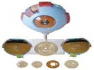
- Product name:6x Magnified Eyeball Anatomy Model (with Base)
- Product code:CC240735
Out of stock.
Request a Quote
Packaging Specifications
Core Parameters
Description
6X Magnified Giant Eyeball Anatomy Model
Detachable eyeball specimen enlarged to six times real size, fitted with supporting stand; structural breakdown as below:
- Outer fibrous tunic: cornea + sclera with extraocular muscle attachments and optic nerve root;
- Middle vascular tunic: iris, ciliary body and choroid;
- Inner nervous tunic: retina;
- Refractive media: crystalline lens and vitreous body.
no comments