Product info
- Product name:Ophthalmic OCT Optical Coherence Tomography Scanner
- Product code:C260127003
Packaging Specifications
- Outer Packaging Size:0.86 × 0.69 × 0.96 m
- Gross Weight:61 kg
- Net Weight:0 kg
- Volume:0.57
- Outer Packaging Size:1.15 × 0.7 × 0.48 m
- Gross Weight:65 kg
- Net Weight:0 kg
- Volume:0.39
- Outer Packaging Size:0.6 × 0.6 × 0.6 m
- Gross Weight:24 kg
- Net Weight:0 kg
- Volume:0.22
Description
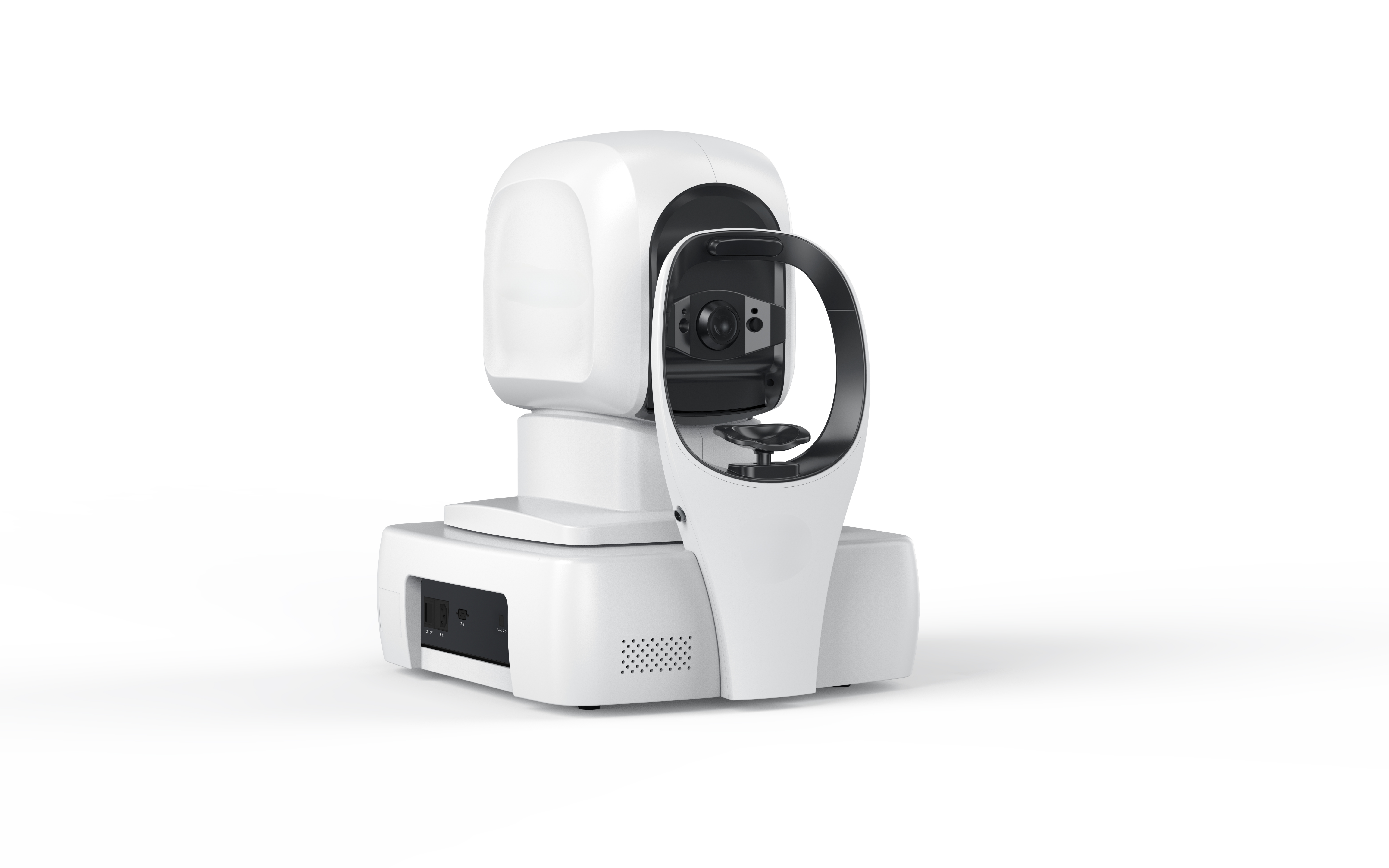
Ophthalmic OCT Optical Coherence Tomography Scanner
New generation fully automated artificial intelligence OCT
OCT-1000 ophthalmic optical coherence tomography (OCT) is a fully automatic artificial intelligence OCT with fully independent intellectual property rights. Combined with multi-image registration and image enhancement technology and industry-leading analysis technology, it provides a full-stack solution for clinical diagnosis and treatment, including automatic image capture, accurate image analysis, intelligent eye health record management, and real-time data sharing on the cloud.
Product Features
Fully automatic operation
High-resolution imaging
Fast pupil positioning and focusing: Full voice guidance; It takes an average of 2 minutes to complete the examination of both eyes of one patient.
12mmX9mm Large field scan; It covers both the macular area and the optic disc area. Retinal disorders can be comprehensively detected
AI Image Analysis
Industry-leading image algorithms
It can be used for auxiliary diagnosis of 16 main fundus lesions. The lesion area was accurately identified, segmented and automatically marked.
Provide reliable and accurate quantitative analysis(such as mean choroidal thickness,central macular thickness, etc.)
Intelligent eye health management
Perfect adaptation to all kinds of scenarios
Automatically establish eye health records for users; Mobile scanning code can quickly receive the inspection report.
Using cloud architecture; Real-time sharing of examination image data; To provide platform support for remote hierarchical medical alliance
Ultra-fast fundus signal acquisition
80,000 times per second "second" acceleration technology, the scanning speed is greatly improved, helping to accurately capture different subdivision forms of fundus lesions in a shorter time and obtain high-quality images.
Hyperfine fundus structure imaging
The AI multimodal ophthalmic image analysis algorithm based on deep learning can more finely observe the subdivision morphology of various fundus diseases and the development of lesions in different structural layers of the retina, realize the hierarchical imaging of the retina and choroid, support the monitoring, diagnosis and hierarchical analysis of the disease, and help clinicians find more disease evidence for patients and guide treatment.
Layering imaging of the retina and choroid in automatic layering mode
Fundus retinal choroidal thickness can be quantified
Ultra-portable and humanized design
The ergonomic fully automatic soft and hard artificial intelligence OCT brings users a new fully automatic equipment experience.
It adopts the split design of optical host and display, combining excellent optical design, automatic algorithm, parallel high-speed computing framework and high ease of use human-computer interaction interface. The imaging quality, analysis accuracy, calculation speed and operation convenience are greatly improved.
Glaucoma analysis function
Disc area
Software analysis
Cup to disc ratio
Horizontal cup to disc ratio
Vertical cup to plate ratio
Optic cup area
RNFL rim sweep thickness curve
RNFL thickness map/TSNum
Topographic map of RNFL thickness
Grid map of TSNI thickness of the nerv fiber layer
66mm fundus map of the optic discThe macular areaSoftware analysisAverage thickness of the inner retinaAverage retinal thicknessMacular foveal thicknessILM-RPE thickness topographyGrid map of ILM-RPE thicknessRetinal volume ETDRS0.01mmTopographic map of RNFL thickness Retinal volume grid map66mm fundus map of the macula
Anterior segment analysis function
High definition single line scan of anterior segment
Six radial lines scan of anterior segment
Diagram of the lesion
It can be used for auxiliary diagnosis of 16 main fundus diseases
1 Retinal Pigment Epithelium Detachment
2 Posterior Vitreous Detachment
3 Epiretinal Membrane
4 Subretinal Fluid
5 Choroidal Neovascularization
6 Drusen
7 Retinoschisis
8 Cystoid Macular Edema
9 Exudation
10 Macular Hole
11 Retinal Detachment
12 Ametropia
13 Elliptical Band Missing
14 Choroidal Excavation
15 Choroidal Atrophy
16 Retinal Hemorrhage
Macular Hole
Macular hole is a tissue loss of the entire layer or part of the macular nerve retina. Most of them are idiopathic and related to abnormal vitreomacular traction, and a few are related to trauma. Lamellar macular hole (LMHH) is a layer of tissue loss on the surface of the fovea and is often associated with the epiretinal membrane.
Cystoid Macular Edema
Posterior Vitreous Detachment
Subretinal Fluid
On OCT, there was a serous detachment under the retinal neuroepithelial layer, with uniform hyporeflection within the detachment area, and a continuous light reflection zone in the retinal pigment epithelial layer below.
Retinal Pigment Epithelium Detachment
Technical parameters
Axial resolution (in the tissue): 5μm
Horizontal resolution (in the tissue): 20um
The highest scanning speed: ≥20000/50000/80000 times/second
Maximum scan range: 12mm*9mm
Centre wavelength: 840nm
Refractive compensation range: -20D ~ + 25D
Scanning modes
Large field: 9mm, Per 30°
Glaucoma: 6mm x 6mm
Single line: Horizontal, Per 30°
Note: When selecting the anterior segment mode,the peripheral devices of the anterior segment should be in place; the design and parameters are subject to change without prior notice.
no comments