❮
❯
Product info
- Product name:Lumbar puncture training model
- Product code:RQK7VA42QV
Packaging Specifications
- Outer Packaging Size:0.99 × 0.59 × 0.34 m
- Gross Weight:17 kg
- Net Weight: kg
- Volume:0.2
Description
Lumbar Puncture & Spinal Anesthesia Training Model
Functional Parameters
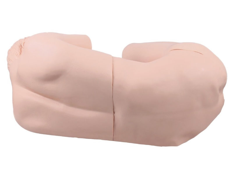
- The simulated standardized patient is placed in the lateral decubitus position, with the back perpendicular to the bed surface, head flexed forward toward the chest, knees flexed toward the abdomen, and the trunk arched.
- The waist is mobile. The operator must hold the simulated patient’s head with one hand and hug the popliteal fossa of both lower limbs with the other to maximize spinal lordosis and widen the intervertebral space before puncture can be performed.
- Accurate lumbar tissue structure and clear surface landmarks: complete L1-L5 vertebrae (vertebral body, lamina, spinous process), sacrum, sacral hiatus, sacral horn, supraspinous ligament, interspinous ligament, ligamentum flavum, dura mater, and arachnoid mater; as well as the subarachnoid space, epidural space, and sacral canal formed by these structures. The posterior superior iliac spine, iliac crest, thoracic spinous processes, and lumbar spinous processes are all truly palpable.
- Supported procedures: spinal anesthesia, lumbar puncture, epidural block, caudal nerve block, sacral nerve block, and lumbar sympathetic nerve block.
- Realistic lumbar puncture simulation: When the puncture needle reaches the simulated ligamentum flavum, resistance increases with a resilient feel. Breaking through the ligamentum flavum produces a distinct "pop" sensation, entering the epidural space with negative pressure (injecting anesthetic at this point simulates epidural anesthesia). Advancing the needle further will puncture the dura mater and arachnoid mater, producing a second "pop" sensation as it enters the subarachnoid space, with simulated cerebrospinal fluid outflow. The entire process simulates the real clinical lumbar puncture procedure.
no comments