Product info
- Product name:5KW 100mA Othopeadic C-arm Image intensifier
- Product code:CCX112B
Packaging Specifications
- Outer Packaging Size:2.5 × 1.1 × 1.48 m
- Gross Weight:575 kg
- Net Weight:335 kg
- Volume:4.07
Description
CCX112B High Frequency Mobile Digital C-arm System
Core Overview
CCX112B is a high-frequency mobile digital C-arm system launched by China Care Medical, featuring a compact and lightweight design, flexible mechanical movement, and low-radiation high-quality imaging performance. Equipped with imported Toshiba core imaging components, a 5KW high-frequency generator, and a professional digital workstation with DICOM3.0 compatibility, it integrates fluoroscopy, photography and digital image processing functions, with humanized operation designs such as electric auxiliary support arm and hand-held controller. Suitable for emergency, general surgery, orthopedics and other clinical departments and surgical scenarios, it provides stable, efficient and safe imaging support for fixation, implant and other surgeries, and can be seamlessly docked with hospital information systems.
Basic Product Parameters
- Packing: 2500x1100x1480mm, total volume 4.07 m³
- Weight: Gross weight 575kg, Net weight 440kg
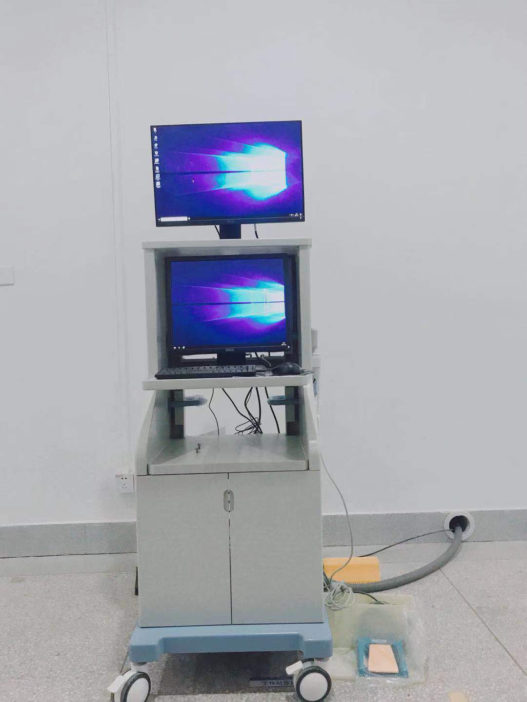
- Core Configuration: Electric auxiliary support arm C-arm frame, 5KW high-frequency HV generator, Toshiba 9-inch three-view image intensifier, imported ultra-low-light digital camera, 19+24 inch LCD displays, DICOM3.0 digital workstation, etc.
Key Technical Specifications
1. Electrical Performance & Exposure Capacity
Fluoroscopy Capacity
- Automatic/Manual Fluoroscopy: Tube voltage 40kV~120kV (auto-adjust), tube current 0.3mA~4mA (manual set)
- Pulse Fluoroscopy: Tube current 0.3mA~30mA (continuous), tube voltage 40kV~120kV
- Max rated capacity: 5KW, inverter frequency 40KHz
Photography Capacity
- Tube voltage 40kV~120kV, tube current 25mA~100mA
- mAs range by voltage segment: 40-49kV(1~180mAs) / 50-59kV(1~140mAs) / 60-69kV(1~125mAs) / 70-79kV(1~110mAs) / 80-89kV(1~100mAs) / 90-99kV(1~80mAs) / 100-110kV(1~63mAs) / 110-120kV(1~50mAs)
2. Core Imaging Components
- X-ray Tube: Fixed anode, dual focus (1.8mm large/0.6mm small); anode capacity 35.5KJ (47KHU), tube thermal capacity 650kJ(867kHu)
- Image Intensifier: Toshiba 9-inch E5764SD-P3, three view (9/6/4.5 inch), image definition 12 bit
- Video System: Japanese imported mega ultra-low-light CCD camera (1024x1024)
- Display: 1 set of 19 inch + 24 inch LCD displays
- Accessories: Dense grain grids, electric adjustable beam collimator, red-cross positioning device
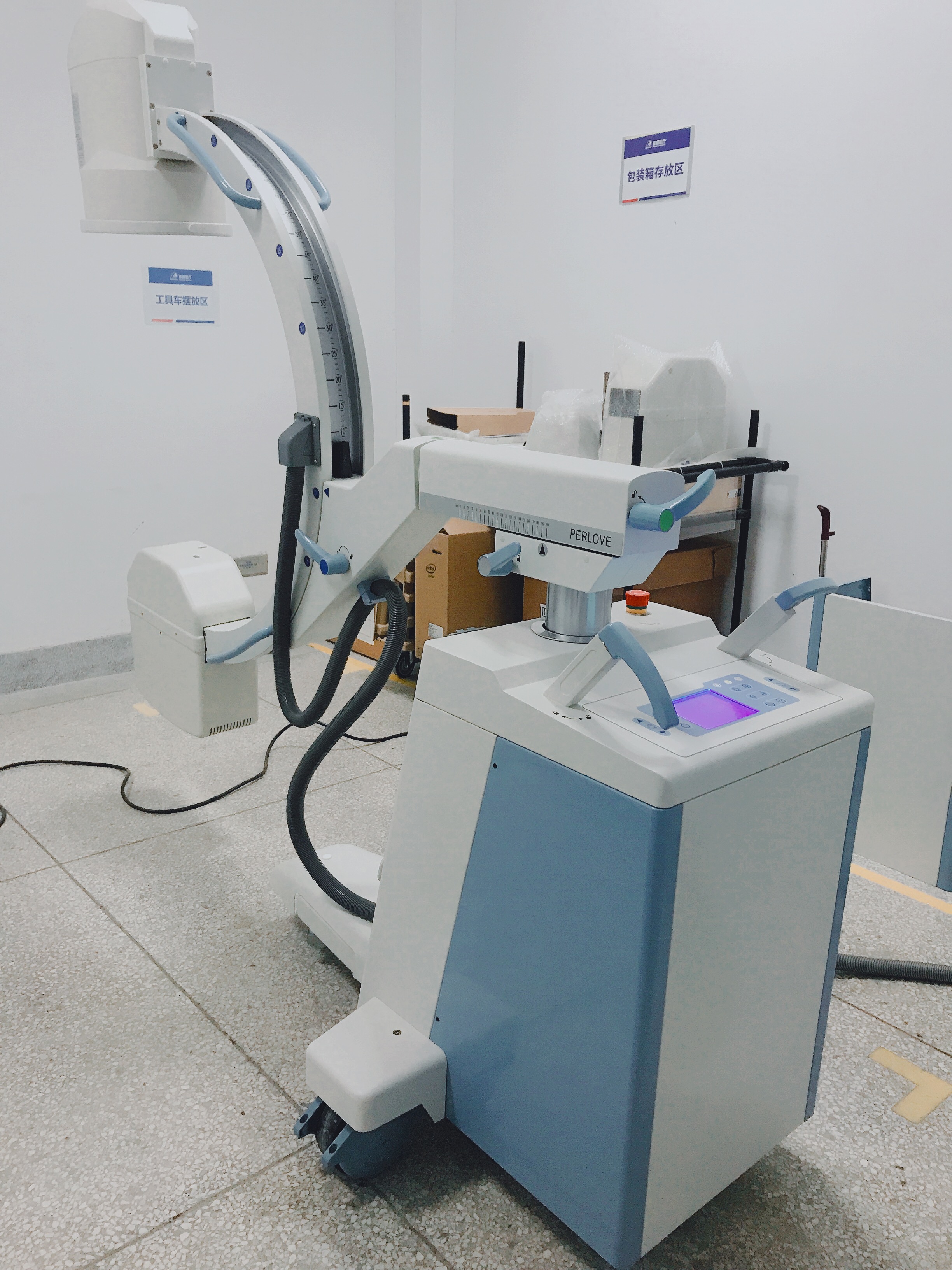
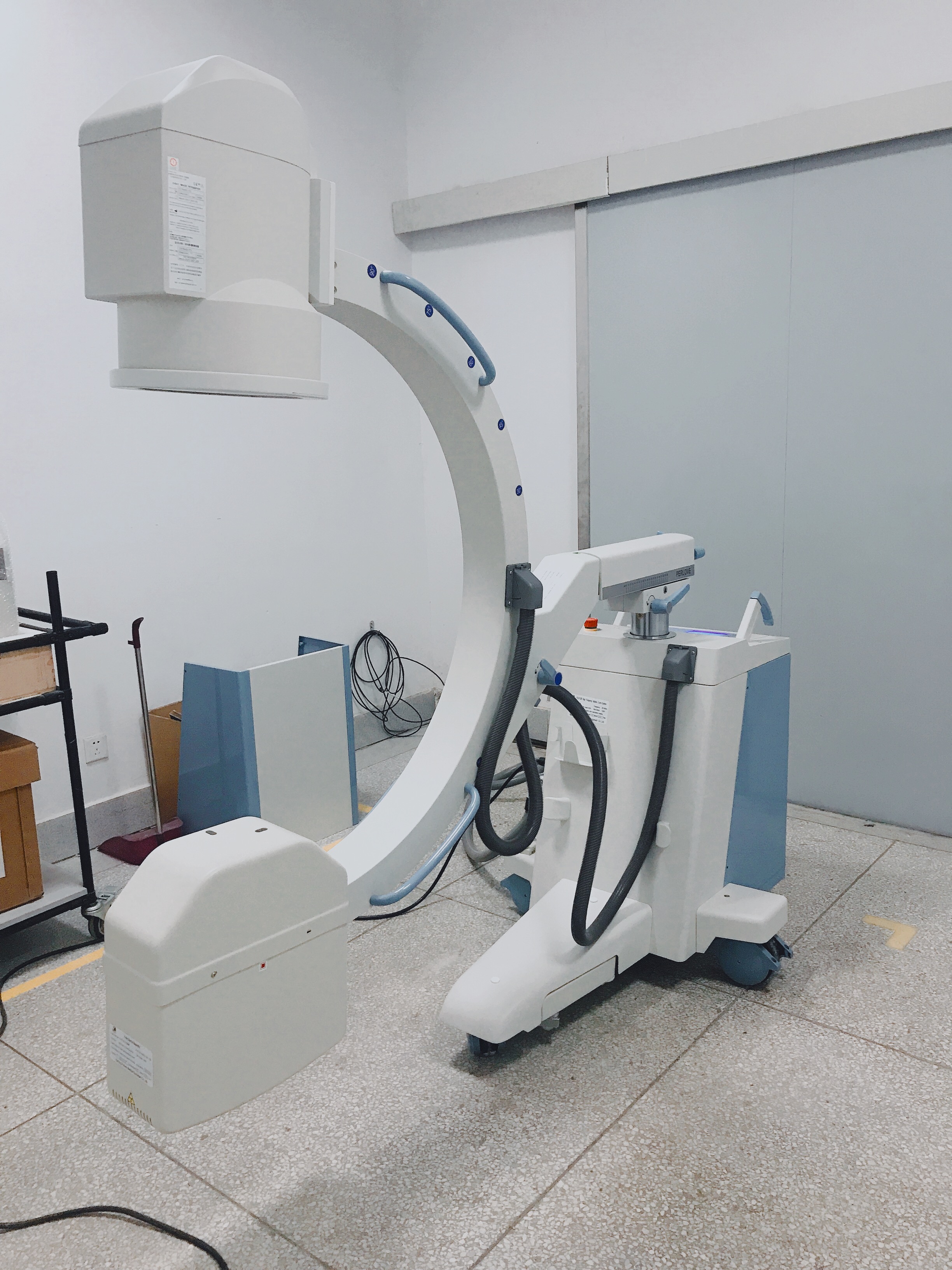
3. Mechanical Structure & C-arm Movement
- Base & Wheels: Directive wheel (360° free direction change); electric auxiliary support arm for stable movement
- C-arm Manual Movement: Revolution around horizontal axis ±180° / swinging ±15° / slip on orbit 120°(+90°~-30°) / forward & backward 200mm
- Motorized Movement: Vertical movement ≥400mm
- Fixed Parameters: SID 1000mm, C-arm opening 760mm, C-arm depth 670mm, directive wheel revolution ±90°
Core Product Advantages
- Compact & Safe Structural Design Lightweight and compact appearance, easy to move and operate, suitable for various operating room spaces Unique base electric auxiliary support arm: Improves the safety and stability of equipment movement and use Diversified movement modes (motorized + manual) and flexible wheel design, realizing all-angle C-arm rotation and free direction adjustment
- Low-Radiation & High-Quality Imaging 5KW high-frequency high-voltage generator with high-precision digital pulse control technology, ensuring excellent images with low skin dose KV/MA automatic tracking function: Dynamically optimizes image brightness and clarity, adapting to different anatomical parts Imported Toshiba three-view image intensifier + ultra-low-light CCD camera, matching dense grain grids, effectively reducing scattered rays and improving image definition Automatic dynamic motion detection: Avoids image blurring caused by patient movement during fluoroscopy
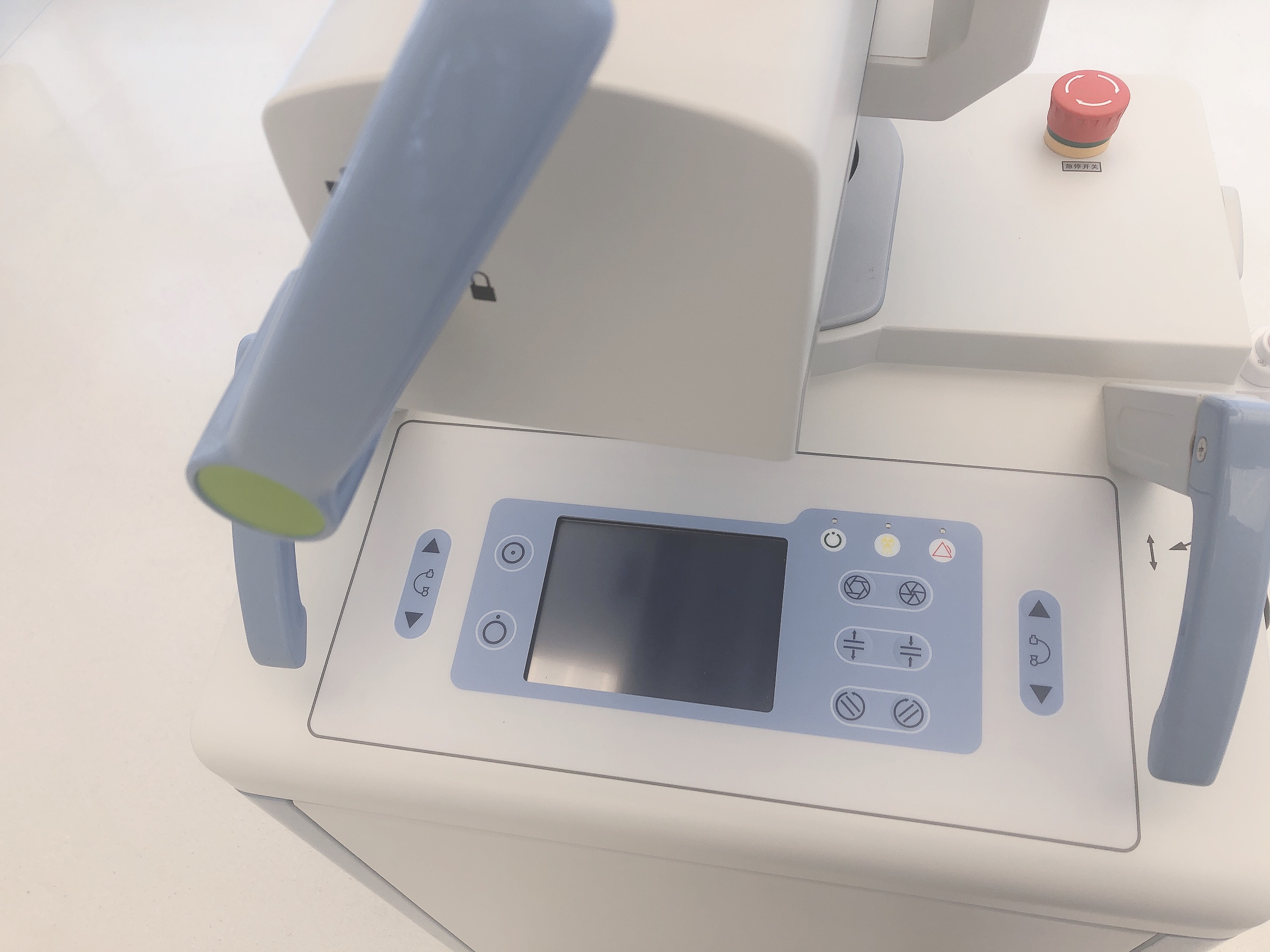
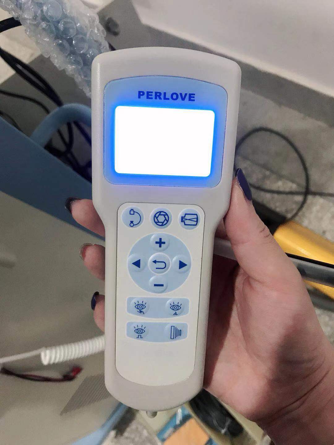
- Humanized & Efficient Operation Unique hand-held controller: Supports long-distance exposure and equipment operation, optimizing surgical workflow Human graphic LCD touch screen system, simple and intuitive parameter setting; foot switch for exposure, freeing hands for surgery Electric controlled collimator, meeting multi-angle and multi-directional anatomical display requirements for different surgeries Red-cross positioning device, realizing accurate lesion positioning and reducing repeated exposure
- Powerful Digital Workstation & System Compatibility Integrated image registration, collection, processing and report output functions: supports window adjustment, edge enhancement, noise reduction, length measurement, expert report templates, etc. Complete DICOM3.0 compatibility: With DICOM browsing, web service and standard interface, seamless docking with hospital PACS, OR, HIS systems and dry film printers Digital radiography function, abandoning traditional film-screen system, simplifying operation processes and realizing digital image storage and sharing
Core Clinical Applications
Suitable for multiple clinical departments and surgical scenarios including Emergency Department, General Surgery, Spine Surgery, Orthopedics, Plastic Surgery and Operating Room; mainly applicable for fixation surgery, implant surgery, and various fluoroscopy-guided interventional operations, meeting the imaging needs of orthopedic reduction, spinal surgery, soft tissue implantation and emergency traumatic surgery.
no comments