Product info
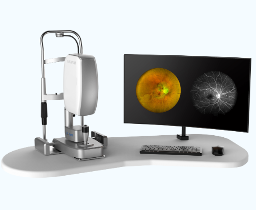
- Product name:Ultra-wide-angle laser fundus angiography with Fundus Autofluorescence FAF and Fundus Fluorescein Angiography
- Product code:CC25042903
Packaging Specifications
- Outer Packaging Size:0.85 × 0.75 × 0.7 m
- Gross Weight:55 kg
- Net Weight:50 kg
- Volume:0.45
- Outer Packaging Size:0.85 × 0.75 × 0.58 m
- Gross Weight:26 kg
- Net Weight:24 kg
- Volume:0.37
- Outer Packaging Size:1.23 × 0.7 × 0.3 m
- Gross Weight:57 kg
- Net Weight:55 kg
- Volume:0.26
Description
CC25042903 / CC25042904 Ultra‑wide‑angle Laser Fundus Angiography System Summary
Manufacturer: China Care Medical Equipment Co.,Ltd
Address: Room B101, ZhongBang Business Building, PuXing Road No. 87, Tianhe, Guangzhou, China
Contact: +86 189 9883 2732;info@chinacaremedical.com;www.chinacaremedical.com
Product Overview
This scanning‑laser‑based ultra‑wide‑field fundus imaging system supports multi‑modal imaging: RGB laser imaging, fundus autofluorescence (Blue AF & Green AF), fluorescein angiography (FFA) and indocyanine green angiography (ICGA). It can stitch 2‑9 captured frames via precise joystick control to generate panoramic images with coverage up to 240°. It supports non‑mydriatic imaging with a minimum pupil size of 2 mm. Confocal laser technology can penetrate cataract and other ocular opacities under poor‑light conditions.
Acquisition Modes & Field‑of‑view Options
- Imaging modes: MCOLOR, IR, RF, AF, ICGA, FFA, ICGA+FFA, Blood Vessel & Cones
- Three FOV grades: 45° UHD, 90° HD, 135° WA
Core Features
- Imaging functions
‑ Live infrared (IR) preview for convenient image acquisition.
‑ Lossless optical zoom; one‑click FOV switching to focus on regions of interest. - Offline Retina Assistant Software (included)
‑ Offline operation without internet access, unlimited license.
‑ Auto‑detection for 20 kinds of ocular pathologies.
‑ Functional tools: image comparison, measurement, annotation and export. - Cell and Blood Flow Tool
‑ Cone‑cell density & average cone‑cell diameter measurement
‑ Red‑blood‑cell flow‑velocity analysis
‑ Vascular wall thickness and diameter measurement - DICOM Compatibility
Built‑in DICOM protocol and worklist support for seamless integration with existing medical imaging systems.
Clinical Imaging Principles
‑ RGB ultra‑wide‑field: three confocal laser sources for high fidelity and high‑contrast fundus images.
‑ Fundus Autofluorescence (FAF): non‑invasive examination for retinal pigment epithelial (RPE) function evaluation, including Blue‑AF and Green‑AF.
‑ FFA: visualizes retinal vasculature with sodium fluorescein dye.
‑ ICGA: long‑wavelength imaging that penetrates RPE to visualize choroidal vessels.
no comments