❮
❯
Product info
- Product name:Orbital magnified anatomical model
- Product code:9MK11UHDBT
Packaging Specifications
- Outer Packaging Size:0.64 × 0.43 × 0.49 m
- Gross Weight:1.1 kg
- Net Weight:14.2 kg
- Volume:0.13
Description
Magnified Orbital Anatomy Model
Product Description
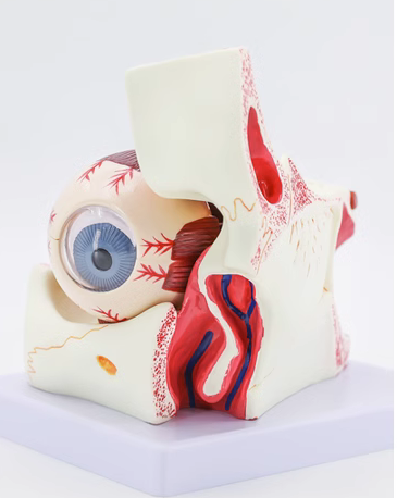
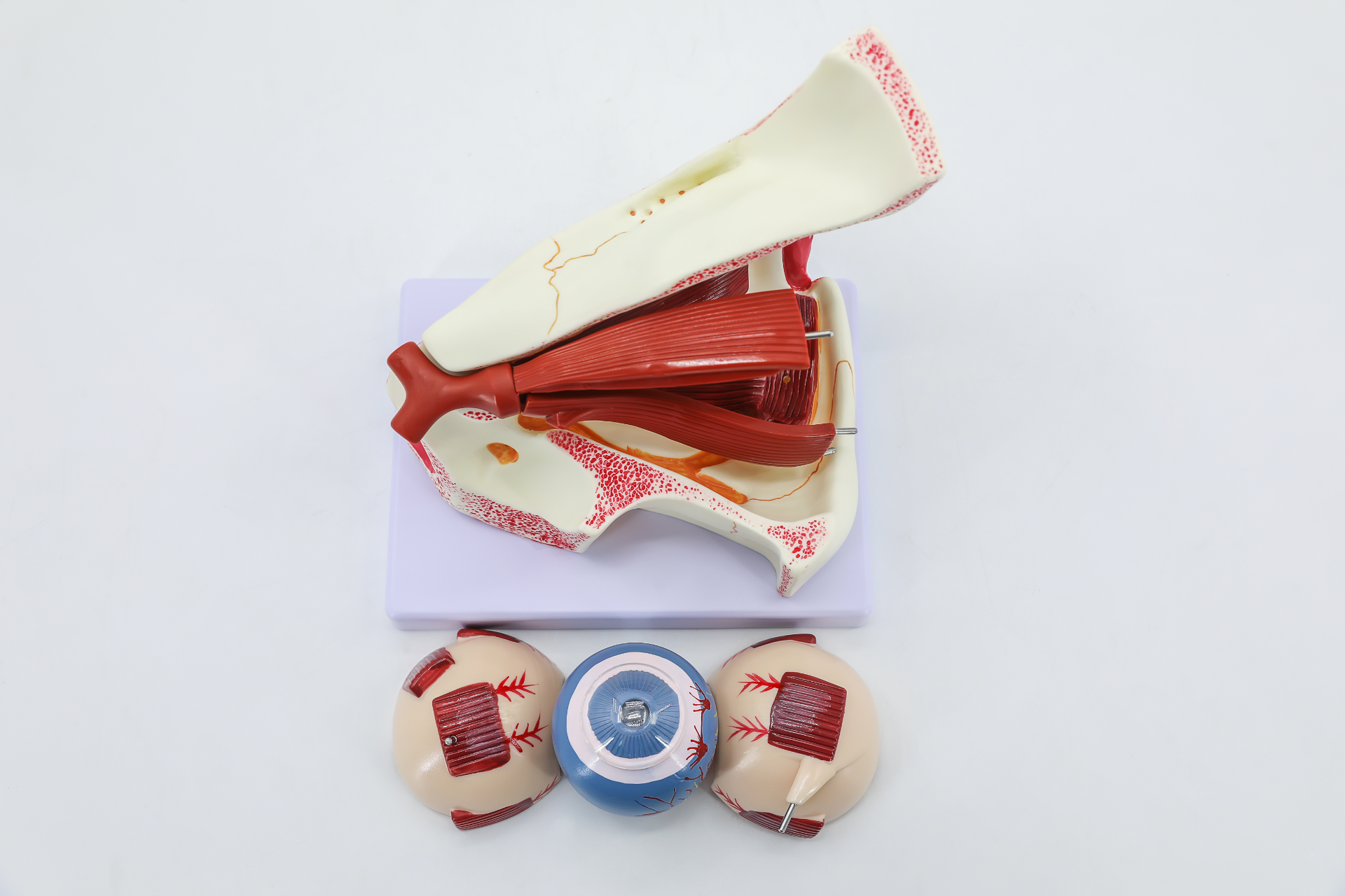
This model is magnified three times the real size. A horizontal cut is made on the eyeball above the maxilla. It is assembled with 10 detachable parts including orbital base, upper and lower sclera segments of eyeball wall, lens, vitreous body, extraocular muscles and optic nerve.
The model exhibits complete eye structures: eyeball wall (sclera, cornea, iris, ciliary body, choroid and retina), intraocular contents, extraocular muscles, accessory eye organs, blood vessels and nerves.
no comments