❮
❯
Product info
- Product name:Advanced 6x Eyepiece (with logo)
- Product code:6NU41GSNYE
Packaging Specifications
- Outer Packaging Size:0.52 × 0.5 × 0.53 m
- Gross Weight:1.55 kg
- Net Weight:13.4 kg
- Volume:0.14
Description
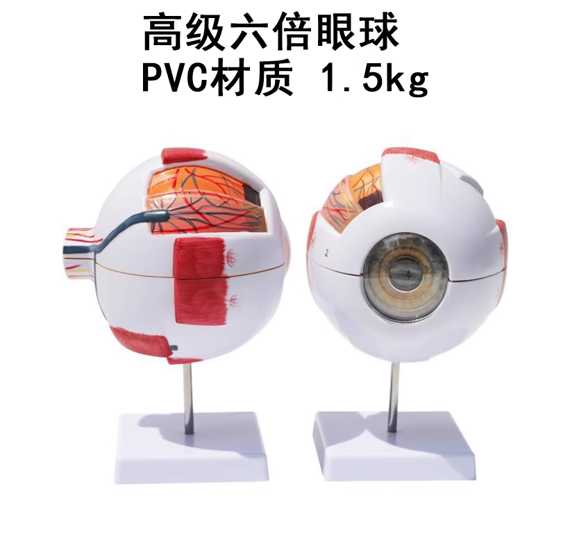
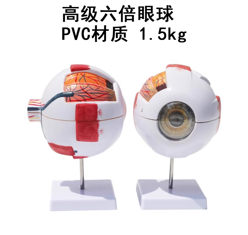
6X Magnified Human Eyeball Anatomy Model
Product Description
This model magnifies human eyeball six times and can be disassembled into six parts, equipped with a display base. The eyeball wall is horizontally cut through the anterior and posterior poles, consisting of three layers: fibrous tunic, vascular tunic and retina.
The crystalline lens and vitreous body are detachable to show the internal macular area and optic nerve structure of eyeball.
Marked with digital labels and matched text explanations, it serves as an intuitive teaching aid for middle schools, colleges and medical universities to illustrate eyeball structure in human anatomy courses.
no comments