Product info
- Product name:85cm male and female torso exchange model (40 parts)
- Product code:M0P8OM2KED
Packaging Specifications
- Outer Packaging Size:0.79 × 0.44 × 0.25 m
- Gross Weight:11.65 kg
- Net Weight:12 kg
- Volume:0.09
Description
85cm Bisexual Interchangeable Torso Anatomy Model (40 Detachable Components)
Product Description
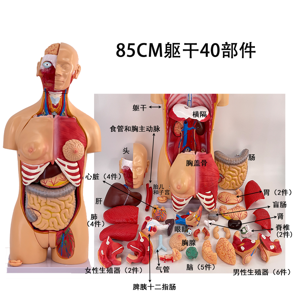
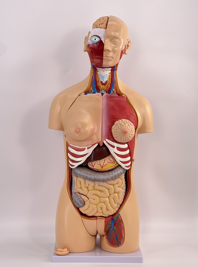
This model illustrates the anatomical positions of human internal organs, as well as the morphological and structural features of the head, showcasing the major human anatomical systems including the respiratory, digestive, urinary, and reproductive systems.
The right half of the head and neck reveals structures such as the skull, masseter muscle, and temporalis muscle, with an eyeball placed within the orbit. A sagittal section is performed on the head and neck: the cranial cavity houses the right cerebral hemisphere, and twelve pairs of cranial nerves are presented on the ventral surface of the brain. It also displays the nasal cavity, oral cavity, and larynx, including the laryngeal cavity, laryngeal ventricle, and rima glottidis. The parathyroid glands are shown posterior to the lateral lobe of the thyroid gland.
The two lungs in the thoracic cavity are subjected to frontal section to reveal the internal structures of the lungs. The heart is coronally dissected to demonstrate the structural differences between the left and right atrioventricular chambers. Cardiac blood vessels including the superior and inferior vena cava, pulmonary artery, pulmonary vein, and aorta are clearly marked for the teaching of systemic and pulmonary circulation.
Full List of Detachable Components
| No. | Component Name | Quantity |
|---|---|---|
| 1 | Main Torso | 1 |
| 2 | Thoracic Cover | 1 |
| 3 | Breast | 1 |
| 4 | Head | 1 |
| 5 | Brain | 5 |
| 6 | Artery Assembly | 1 |
| 7 | Eyeball | 1 |
| 8 | Lung | 4 |
| 9 | Trachea and Bronchus | 1 |
| 10 | Esophagus | 1 |
| 11 | Heart | 4 |
| 12 | Liver | 1 |
| 13 | Sectioned Kidney | 1 |
| 14 | Pancreas, Spleen and Duodenum Complex | 1 |
| 15 | Stomach | 2 |
| 16 | Large Intestine | 1 |
| 17 | Sectioned Cecum | 1 |
| 18 | Interchangeable Male and Female Reproductive Organs | 8 |
| 19 | 12th Thoracic Vertebra with Spinal Cord | 2 |
| 20 | Diaphragm | 1 |
| 21 | Embryo | 1 |
| Total | - | 40 |
no comments