 USD
USD EUR
EUR GBP
GBP CFA
CFA
- Description
Specifications
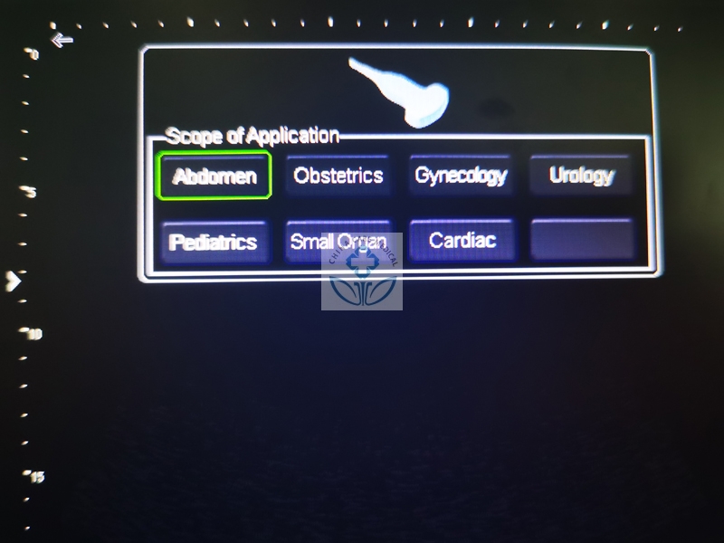
Application:
Abdomen, OB/GYN, Small Parts, MSK, Vascular, Anaesthesia, Emergency
Style:
Portable
Types:
Black and White
Video:
Standard Configuration:
Main Unit ---1
Convex Probe---1
Aluminum Case---1
Battery---1
Main Parameters
1.Operating system: using ARM chip architecture, stable, concise and powerful
2.Display: 12-inch 1024*768 resolution high-definition LED LCD screen
3.Imaging modes: B, B+B, B+M, M, 4B
Probe array element: automatically identify and use a variety of array element probe(80 elements,96 elements,128 elements)
4.Image preset: The system combines a large amount of clinical experience, has different preset adjustment conditions for the parts or organs, and has a one-key optimization function
5.Cine playback: ≥256 frames
6.Image storage: ≥360 frames.External USB storage,Single image storage time≤about 9s.(Storage speed is related to the USB device used)
7.Scanning angle adjustment: adjustable
8.Image enlargement: Separate knob depth adjustment (≥about 20 levels adjustable)
9.Puncture: with puncture guide line, angle and position are adjustable
10.Focus: more than 5 levels of focus position adjustment
11.Image adjustment: up and down, left and right, brightness, focus number, focus position, dynamic range, scan angle, frame correlation, M speed
12.Image Processing: image smoothing/sharpening, tissue harmonics, gamma correction, false color
13.Notes and characters: date, clock, name, age, gender, doctor, hospital name, image annotation
14.Position mark: ≥97 kinds
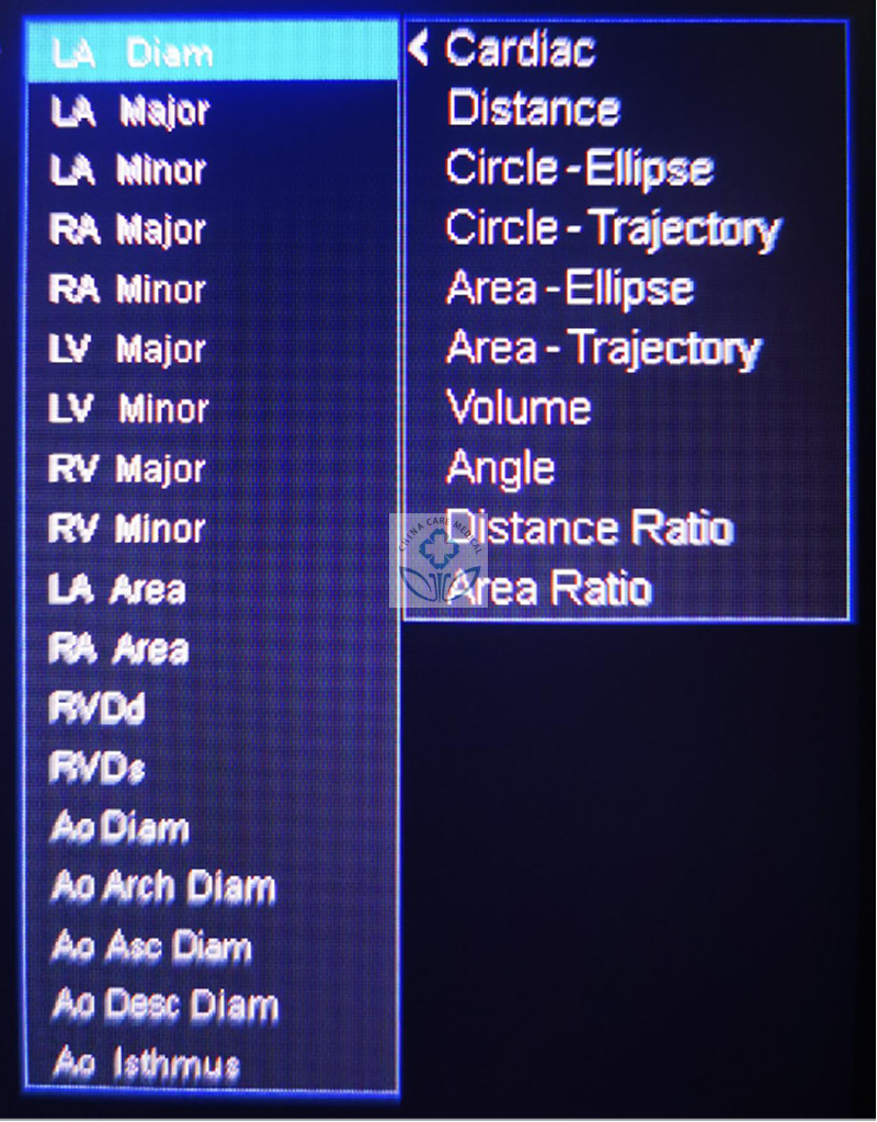
15.Measurement function
a) Routine measurement: distance, circumference, area, volume;
b) Heart measurement: left atrium, right atrium, left ventricle, right ventricle, aorta, descending aorta, aortic isthmus;
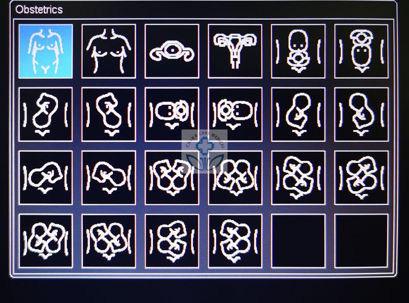
c) Obstetric measurement: gestational sac, biparietal diameter, head circumference, abdominal circumference, femur length, fibula length, head hip diameter, yolk sac, abdomen, cerebrum, cerebellum, amniotic fluid, gestational age, expected date of delivery, fetal weight, etc;
d)Gynecological measurement: uterus, cervix, endometrial thickness, ovaries, follicles;
e)Urology measurement: kidney, adrenal gland, prostate, testicular mass, epididymis, etc;
f)Pediatric measurement: common liver duct, common bile duct, pancreas, spleen, kidney, adrenal gland, etc;
g)Small organ measurement: thyroid, seminal vesicle, testis, scrotum, breast mass, skin mass, etc.
The instrument has different ethnographic measurement formulas.
16.Report function: automatically generate reports on abdomen, urology, obstetrics, heart, etc
17.External interface: VGA interface, USB2.0 interface, RS232 interface
18.Power saving mode: the panel button light can be turned on and off by one button, and the instrument will automatically turn off the power if it is not used for 6 minutes. Press any key to resume operation
19.Language settings: Chinese and English
20.Built-in operating instructions
Usage introduction
First turn on the power switch at the bottom left of the instrument, and then press the POWER key, the instrument automatically enters the operation interface.
First press PROBE (probe selection key) to select department or organ. The instrument pre-sets the conditions of each department or inspection site. Customers can make the following fine-tuning according to their own habits:
1: Slide the TGC left and right to adjust the near, middle and far field signals;
2: Rotate the GAIN knob to adjust the overall image gain.
3: Rotate the DEPTH knob to adjust the depth.
4: Adjust the focus position to adjust the definition of the position to be scanned.
5: Adjust FREQ, the higher the frequency, the more delicate the image, but the less the signal.
6: Adjust dynamic range DR, spot suppression NOI CAN, , the higher the value, the finer the image, but the less the signal richness.
7:Adjust the image signal richness and brightness:press MENU to enter ther settings-grayscale brightness. It is recommended to adjust in the range of 11-16 under different light.
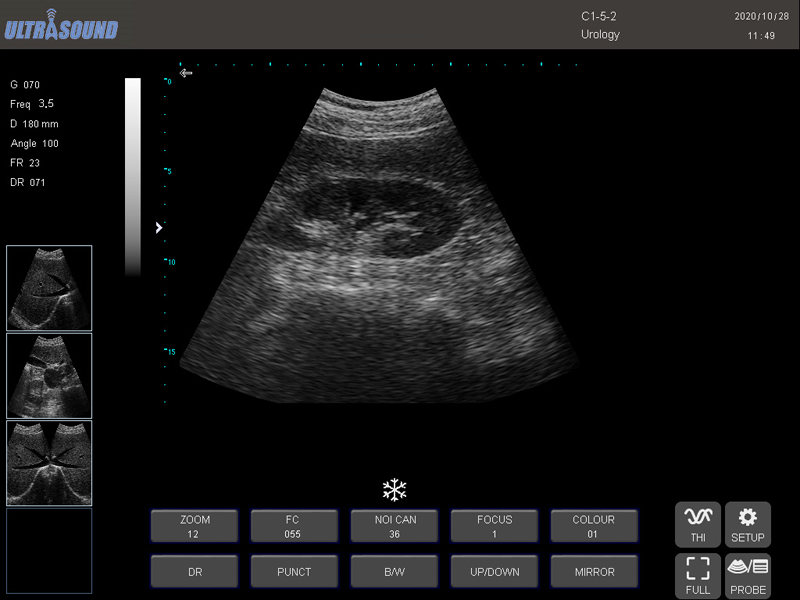
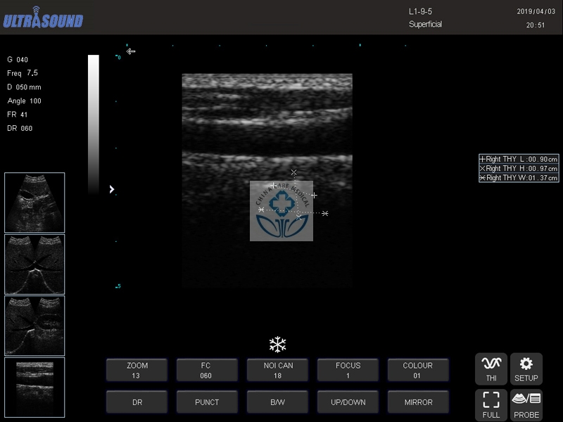
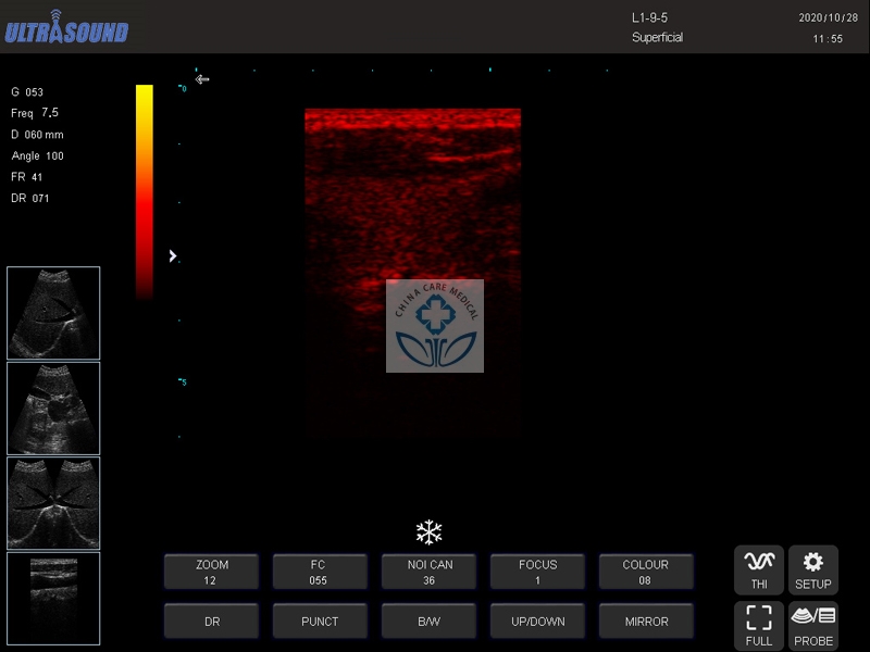
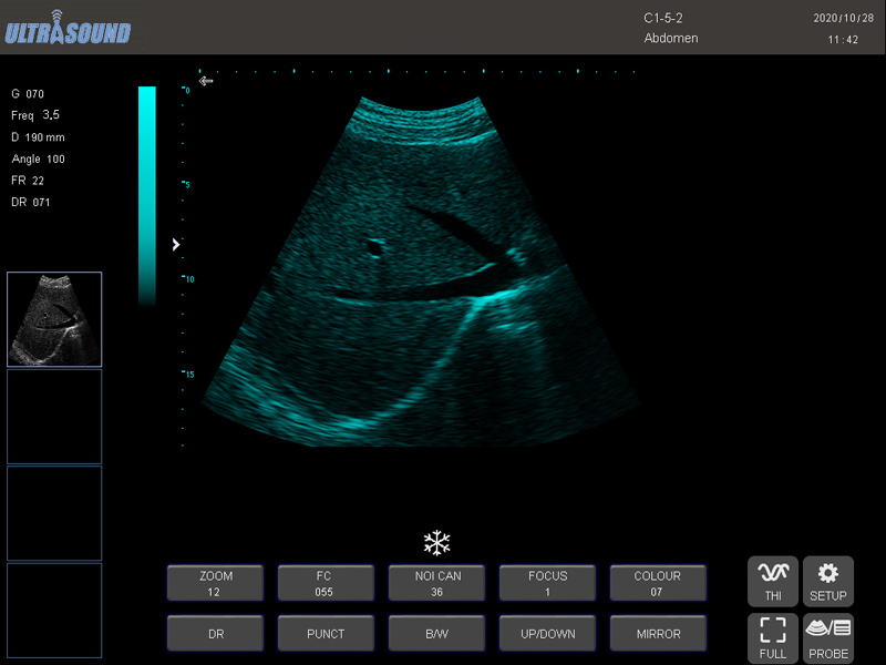
Imagines Pictures:
Operation Pictures:

Patient Report

Standard Configuration:
Main Unit ---1
Convex Probe---1
Aluminum Case---1
Battery---1
Main Parameters
1.Operating system: using ARM chip architecture, stable, concise and powerful
2.Display: 12-inch 1024*768 resolution high-definition LED LCD screen
3.Imaging modes: B, B+B, B+M, M, 4B
Probe array element: automatically identify and use a variety of array element probe(80 elements,96 elements,128 elements)
4.Image preset: The system combines a large amount of clinical experience, has different preset adjustment conditions for the parts or organs, and has a one-key optimization function
5.Cine playback: ≥256 frames
6.Image storage: ≥360 frames.External USB storage,Single image storage time≤about 9s.(Storage speed is related to the USB device used)
7.Scanning angle adjustment: adjustable
8.Image enlargement: Separate knob depth adjustment (≥about 20 levels adjustable)
9.Puncture: with puncture guide line, angle and position are adjustable
10.Focus: more than 5 levels of focus position adjustment
11.Image adjustment: up and down, left and right, brightness, focus number, focus position, dynamic range, scan angle, frame correlation, M speed
12.Image Processing: image smoothing/sharpening, tissue harmonics, gamma correction, false color
13.Notes and characters: date, clock, name, age, gender, doctor, hospital name, image annotation
14.Position mark: ≥97 kinds
15.Measurement function
a) Routine measurement: distance, circumference, area, volume;
b) Heart measurement: left atrium, right atrium, left ventricle, right ventricle, aorta, descending aorta, aortic isthmus;
c) Obstetric measurement: gestational sac, biparietal diameter, head circumference, abdominal circumference, femur length, fibula length, head hip diameter, yolk sac, abdomen, cerebrum, cerebellum, amniotic fluid, gestational age, expected date of delivery, fetal weight, etc;
d)Gynecological measurement: uterus, cervix, endometrial thickness, ovaries, follicles;
e)Urology measurement: kidney, adrenal gland, prostate, testicular mass, epididymis, etc;
f)Pediatric measurement: common liver duct, common bile duct, pancreas, spleen, kidney, adrenal gland, etc;
g)Small organ measurement: thyroid, seminal vesicle, testis, scrotum, breast mass, skin mass, etc.
The instrument has different ethnographic measurement formulas.
16.Report function: automatically generate reports on abdomen, urology, obstetrics, heart, etc
17.External interface: VGA interface, USB2.0 interface, RS232 interface
18.Power saving mode: the panel button light can be turned on and off by one button, and the instrument will automatically turn off the power if it is not used for 6 minutes. Press any key to resume operation
19.Language settings: Chinese and English
20.Built-in operating instructions
Usage introduction
First turn on the power switch at the bottom left of the instrument, and then press the POWER key, the instrument automatically enters the operation interface.
First press PROBE (probe selection key) to select department or organ. The instrument pre-sets the conditions of each department or inspection site. Customers can make the following fine-tuning according to their own habits:
1: Slide the TGC left and right to adjust the near, middle and far field signals;
2: Rotate the GAIN knob to adjust the overall image gain.
3: Rotate the DEPTH knob to adjust the depth.
4: Adjust the focus position to adjust the definition of the position to be scanned.
5: Adjust FREQ, the higher the frequency, the more delicate the image, but the less the signal.
6: Adjust dynamic range DR, spot suppression NOI CAN, , the higher the value, the finer the image, but the less the signal richness.
7:Adjust the image signal richness and brightness:press MENU to enter ther settings-grayscale brightness. It is recommended to adjust in the range of 11-16 under different light.
Imagines Pictures:
Operation Pictures:
Patient Report
Shipping Information:
G.W: 8kg
Packing Size: 0.42m,0.39m,0.19m
Unit: Piece
Special: With Battery
Name:
Email:
Whatsapp/Tel:
Message:
You May Like

-
$2,860.00

-
$3,250.00

-
$5.00

-
$7,098.00

-
$3,978.00

-
$100.00

-
$3.00

-
$0.35

-
$14.00